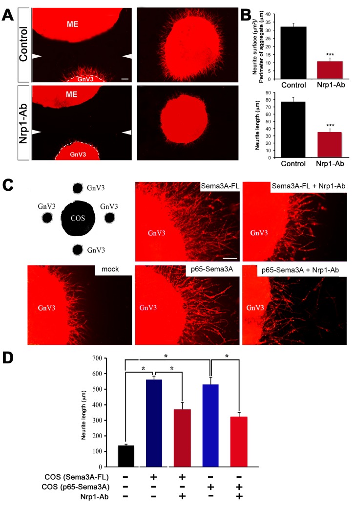
Figure 5. 65-Nrp1 signaling is responsible for GnRH neurite sprouting.
(A) Three-dimensional matrix assays using co-cultures of ME explants dissected from adult female rats (control, n = 3; Nrp1-Ab, n = 4) and cell aggregates of immortalized GnV-3 cells, in the absence (top panels) or presence (bottom panels) of an Nrp1-neutralizing antibody (Nrp1-Ab). Co-cultures were fixed and stained with Alexa 588–X phalloidin. GnV-3 cell aggregates show neurite extension under control conditions, whereas neurite sprouting is strongly inhibited by Nrp1-Ab. White arrowheads in the left panels indicate the merging point of the two individual images composing each picture. (B) Quantitative analysis of the area covered by phalloidin staining surrounding the aggregates (top panel; n = 3 in controls, n = 4 in Nrp1-Ab-treated aggregates; unpaired Student's t test, t(5) = 7.424, p<0.001) and GnV-3 neurite length (bottom panel; n = 3 in controls, n = 4 in Nrp1-Ab-treated aggregates; unpaired Student's t test, t(5) = 5.610, p<0.005), respectively. (C) Co-cultures of GnV-3 cell aggregates placed around aggregates of COS-7 cells transfected with full-length (95 kDa) Sema3A (Sema3A-FL), 65 kDa Sema3A (p65-Sema3A), or the control vector (n = 7), in the presence (Sema3A-FL+Nrp1-Ab, n = 4; p65-Sema3A+Nrp1-Ab, n = 6) or absence of Nrp1-Ab (Sema3A-FL, n = 4; p65-Sema3A, n = 4), as shown in the schematic drawing. Sema3A-FL and p65-Sema3A are equally effective at inducing GnV-3 neurite growth when compared to control conditions (middle panels), while neurite growth is prevented by the Nrp1-neutralizing antibody (right panels). (D) Quantitative analysis of GnV-3 neurite length (one-way ANOVA with Tukey's post hoc test, F(4,24) = 38.058, p<0.0001). Data are represented as means ± SEM. Scale bars, 100 µm in (A), 50 µm in (C).

