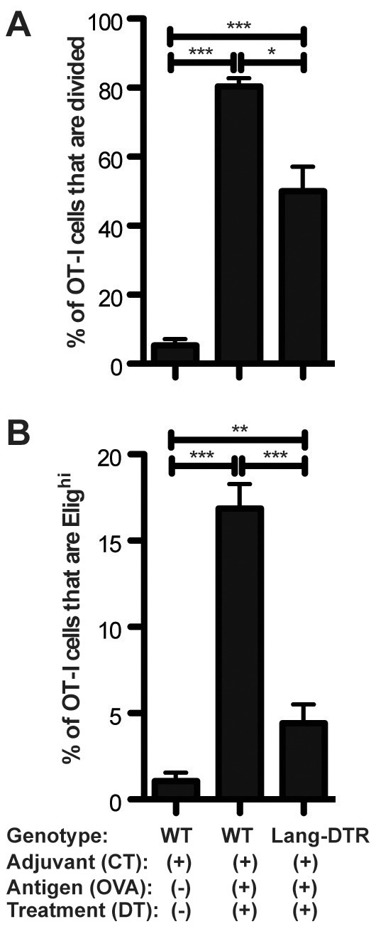
Figure 5. Decreased proliferation and E-lig induction for OT-I cells adoptively transferred into Lang+ DC-depleted mice.

CFSE-labeled splenocytes from CD45.1+ OT-I donor mice were injected iv into recipient WT and Lang-DTR mice. On the following day, recipients were topically immunized with CT+OVA protein or CT alone on the ear skin, using the same immunization techniques as in our in vivo/ex vivo assays. All mice were treated with twice diphtheria toxin. Timeline: day -2, first DTX treatment; day -1, OT-I cells transferred IV to recipients; day 0, ear skin immunized and second DTX treatment given; day 5, skin-draining LN harvested. A: Proliferation depicted as the percentage of total OT-I T cells that are CFSE low. B: E-lig expression depicted as the percentage of total. N = 3 experiments of 4–5 mice per group. For all experiments shown, sdLN cells were isolated and gated on CD45.1+CD3+CD8+ cells. One-tailed Mann-Whitney p values shown. *p<.05; **p<.001; ***p<.0001; n.s. = not significant.
