
Figure 1. Chain disposition of Asterionellopsis glacialis visualized at 200× magnification with an inverted microcope (Leica DMIL).
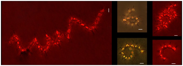
The photographs chosen are representative of chains of different lengths irrespective of the carbon dioxide concentration (photos in red show auto-fluorescence achieved by using the filter N2.1 green). Note the proximity between cells in the spirals. Scale bars correspond to 10 µm.