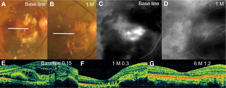
Figure 3.

Findings in a 63-year-old man who underwent pneumatic displacement combined with intravitreal bevacizumab (PD + IVB). (A and B) Fundus photographs. (C and D) Indocyanine green angiograms. (E–G) Optical coherence tomographic images. Visual acuity was 0.15. (A) Photographs of fundus show 4 disc diameter massive submacular hemorrhage in the right eye. Some hemorrhagic retinal pigment epithelium detachments can be seen in the hemorrhage. (B) One month after treatment with PD + IVB, the fundus photograph shows no submacular hemorrhage. Visual acuity was 0.8. (C) Indocyanine green angiograms showing active polypoidal dilatation of choroidal vessels at the site of hemorrhagic retinal pigment epithelium detachments. (D) Indocyanine green angiograms showing disappearance of leakage from the polypoidal choroidal vasculopathy 1 month after treatment. (E) Baseline horizontal optical coherence tomography image showing thick submacular hemorrhage and hemorrhagic pigment epithelium detachments. (F) Horizontal optical coherence tomography image 1 month after treatment with PD + IVB showing a decrease in central retinal thickness. Hemorrhagic retinal pigment epithelium detachments and mild macular edema remain. (G) Horizontal optical coherence tomography image 6 months after treatment with PD + IVB showing a smooth macular image. No exudative changes remain in the macular area.
Abbreviations: IVB, intravitreal bevacizumab; PD, pneumatic displacement; M, month.
