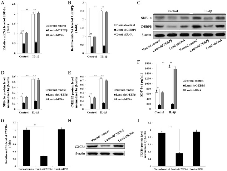
Figure 4. mRNA and protein expression of SDF1α and C/EBP-β by transfected EPCs, analyzed by real-time quantitative RT-PCR, western blotting and ELISA.
Untreated EPCs or EPCs transfected for 12-shC/EBPβ or Lenti-shRNA, were treated with 10 ng/mL IL-1β or control vehicle (DMSO) for 24 hours, then mRNA levels of SDF1α (A) and C/EBP-β (B) were detected by Q RT-PCR. GAPDH served as control. C, protein levels of SDF1α and C/EBP-β were detected by western blot analysis. β-actin served as loading control. Quantitative analysis of SDF1α (D) and C/EBP-β (E) protein levels normalized to β-actin. F, Levels of secreted SDF1α in the medium of the above EPC cultures (Normal EPCs, Lenti-shC/EBPβ, Lenti-shRNA-EPCs) were determined by ELISA. mRNA and protein expression of CXCR4 by transfected RAW264.7 cells, analyzed by real-time quantitative RT-PCR and western blotting. G, Untreated RAW264.7 cells or RAW264.7 cells transfected with Lenti-shCXCR4 or Lenti-shRNA for 48 h, were subjected to Q RT-PCR to measure the mRNA levels of CXCR4. Results were normalized to GAPDH expression. H, Protein levels of CXCR4 were analyzed by Western blot. β-actin served as loading control. I, Quantitative analysis of CXCR4 protein level normalized to β-actin. All values are the means ± SD of three replicates, * P<0.05, ** P<0.01.

