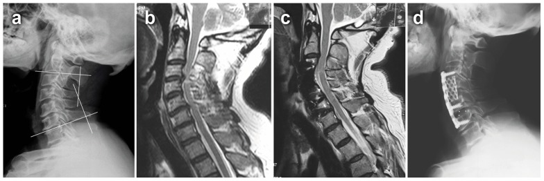
Figure 2. A 65-year-old male developed numbness in his two hands and weakness in his four extremities for 2 years.

Preoperative imaging studies showed that the spinal cord compressed at C3–C6. He was performed with 3-level ACHDF. After operation, his JOA scores improved from 7 preoperation to 13 postoperation. a Preoperative lateral X-ray. The segmental lordosis of C2–C7 was defined as the angle formed by the lower endplate of C2 vertebral body and the upper endplate of C7 vertebral body. b Preoperative MRI. c 2-year postoperative MRI. d 2-year postoperative lateral X-ray.
