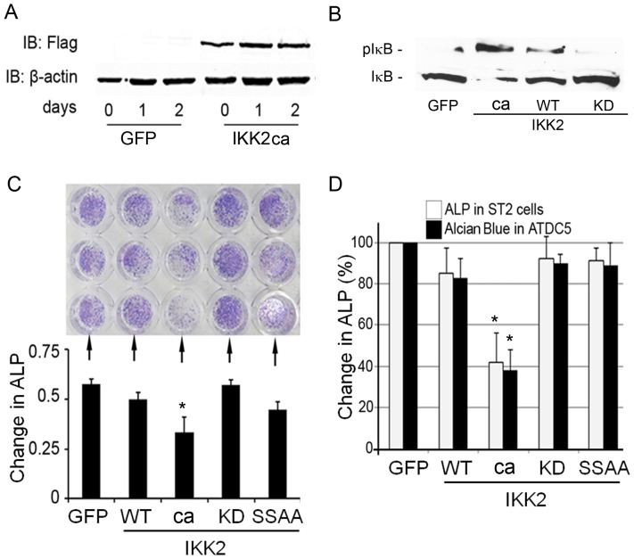
Figure 1. Constitutive activation of NF-κB in osteoblasts, chondrocytes and stromal cells inhibits their differentiation.
(A) Calvarial osteoblasts (cOB) were transduced using retroviral constructs pMX-GFP and pMX-IKK2-ca and incubated for the time points indicated. Western blots were used to show expression of Flag-tagged IKK2 and β-actin in cOB. (B) Expression of IκB and phospho-IκB in lysates of OBs transduced with pMx-GFP or the various forms of IKK2 as indicated. (C–D) Calvarial OBs (C), ST2 and ATDC5 cells (D) were infected with GFP, IKK2WT, IKK2ca, IKK2KD, or IKK2SSAA and incubated for 21 days with 10 ug/ml of bovine insulin (media was changed and supplemented with fresh insulin every 48 hrs). Cells were then stained with either alkaline phosphatase (ALP) or Alcian blue. Note reduced staining in IKK2ca conditions. Lower left panel (C) depicts optical density quantification of upper panel. Right panel (D) represents quantification of ST2 cell and ATDC5 staining (not shown) using arbitrary units expressed as % of control. Experiments were repeated at least three times in triplicate conditions. Asterisk represents p<0.01.

