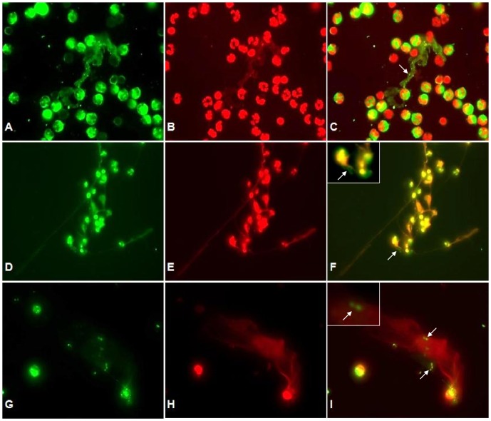
Figure 2. Co-localization of DNA with histones (H3), NE and MPO in tachyzoite-induced NET structures.
Co-cultures of bovine PMN and B. besnoiti tachyzoites were fixed, permeabilized, stained for DNA using Sytox Orange (red: B, E, H) and probed for MPO (green: A), histones (green: D) and NE (green: G) using anti-MPO, anti-histone (H3) and anti-NE antibodies and adequate conjugate systems. Areas of respective co-localization (merges) are illustrated in C, F, I. The arrow in (C) indicates delicate globular structures within NETs. Arrows in (F) and (I) indicate tachyzoites being trapped in NET structures. Photomicrographs are of representative cells from 3 independent experiments. The time culture in this experiment was 60 min.

