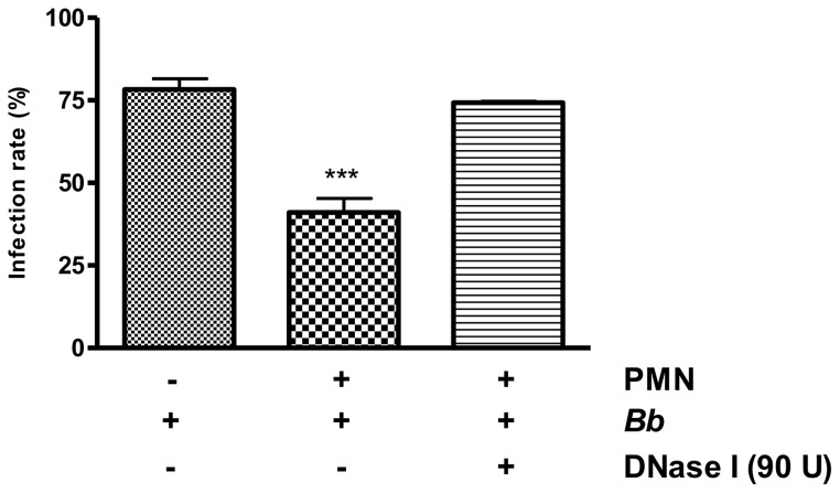
Figure 9. Infectivity of B. besnoiti tachyzoites after exposure to bovine PMN.
Vital B. besnoiti tachyzoites were co-cultured for 3 h with bovine PMN ( = PMN + B.b.) allowing for effective NET formation. To dissolve potential NET structures, DNase was supplemented 15 min before the end of the incubation period ( = B.b. + PMN + DNase). Incubation of tachyzoites in plain medium served as PMN-free, infection control ( = B.b. only). After incubation, samples were transferred to confluent BUVEC monolayers for 1 h. Thereafter, the cell layers were thoroughly washed and infection rates were estimated. Arithmetic means and standard deviations of three PMN donors, minimum and maximum. Differences were regarded as significant at a level of p≤0.05.

