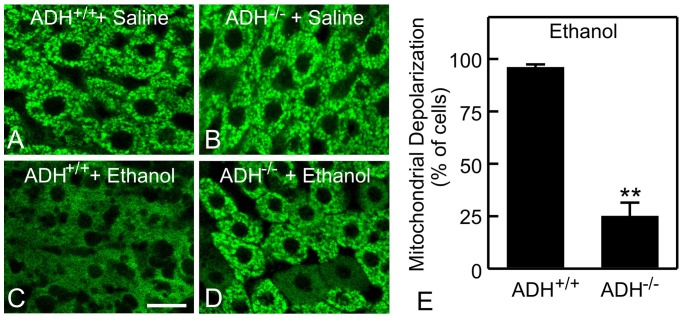
Figure 5. Role of alcohol dehydrogenase in ethanol-induced mitochondrial depolarization.
ADH positive (ADH+/+) and negative (ADH-/-) deer mice were gavaged with saline (A and B) or ethanol (6 g/kg, C and D). Rh123 fluorescence was detected after 6 h. Representative images of 4 mice per group are shown. Bar is 10 µm. E: quantification of cells with mitochondrial depolarization at 6 h after ethanol treatment. Values are means ± SEM (n = 4 per group). **, p<0.01 vs ADH positive deer mice.

