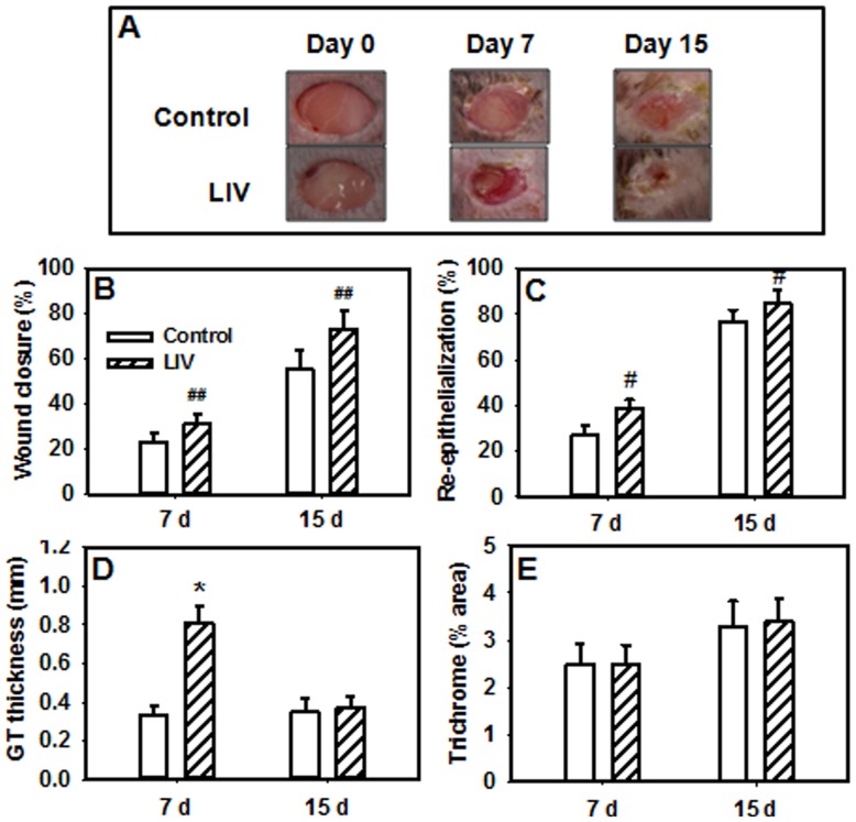
Figure 2. Low-intensity vibration enhances wound healing in db/db mice.
Mice received either low-intensity vibration (LIV; 0.4 g at 45 Hz) or a sham control treatment starting the day of wounding for 5 d/wk. Representative images of wounds were taken on days 0, 7 and 15 post-injury (a). Wound closure was measured in digital images of wound surface, 7 d, n = 12–13 per group; 15 d, n = 3 per group (b). Re-epithelialization was measured in hematoxylin and eosin stained sections of wound center, 7 d, n = 13–16 per group; 15 d, 8–9 per group (c). Granulation tissue thickness was measured as the area of granulation tissue divided by the distance between wound edges, 7 d, n = 13–16 per group; 15 d, 8–9 per group (d). Collagen deposition was measured in trichrome stained sections as the percent area stained blue, 7 d, n = 13–16 per group; 15 d, 8–9 per group (e). #Main effect of treatment, P≤0.05. ##Main effect of treatment, P = 0.066. *Mean value significantly different from that of control for same time point, P≤0.05.

