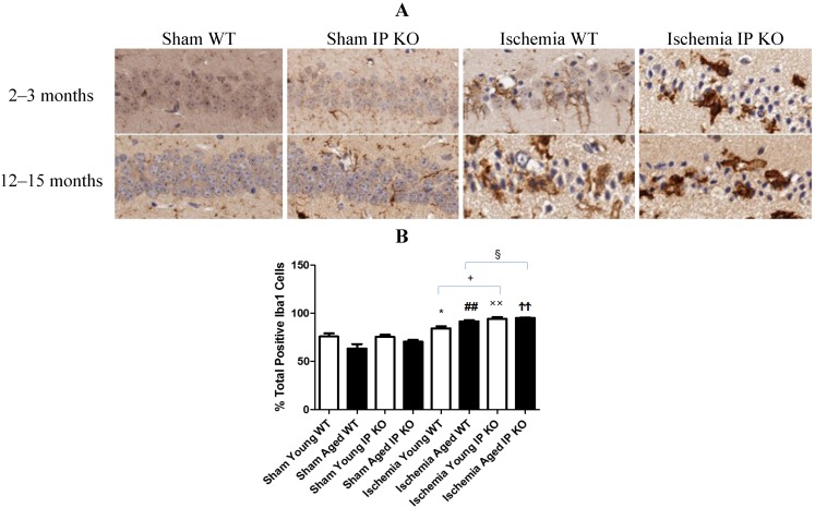
Figure 5.
Microglia reactivity in young and aged WT and IP KO sham and ischemic mice after 12 min global cerebral ischemic insult. (A) Representative images of brain sections show microglia immunostaining. (B) Quantification of Iba-1-positive cells in the hippocampal CA1 subfield on day 7 after ischemia. (n = 3 per group). * p < 0.05, ## p < 0.01, ×× p < 0.01, ϮϮ p < 0.01 for Ischemia vs. Sham; + p < 0.05, § p < 0.05 for Ischemia IP KO vs. Ischemia WT.

