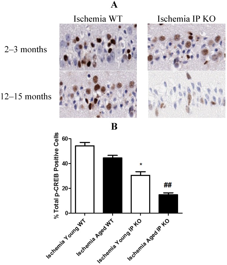
Figure 7.
The phosphorylation of CREB in young and aged WT and IP KO mice after 12 min global cerebral ischemic insult. (A) Representative images of brain sections show p-CREB immunostaining. (B) Quantification of p-CREB positive cells in the hippocampal CA1 subfield on day 7 after ischemia. (n = 3 per group). * p < 0.05, ## p < 0.01 for IP KO vs. WT.

