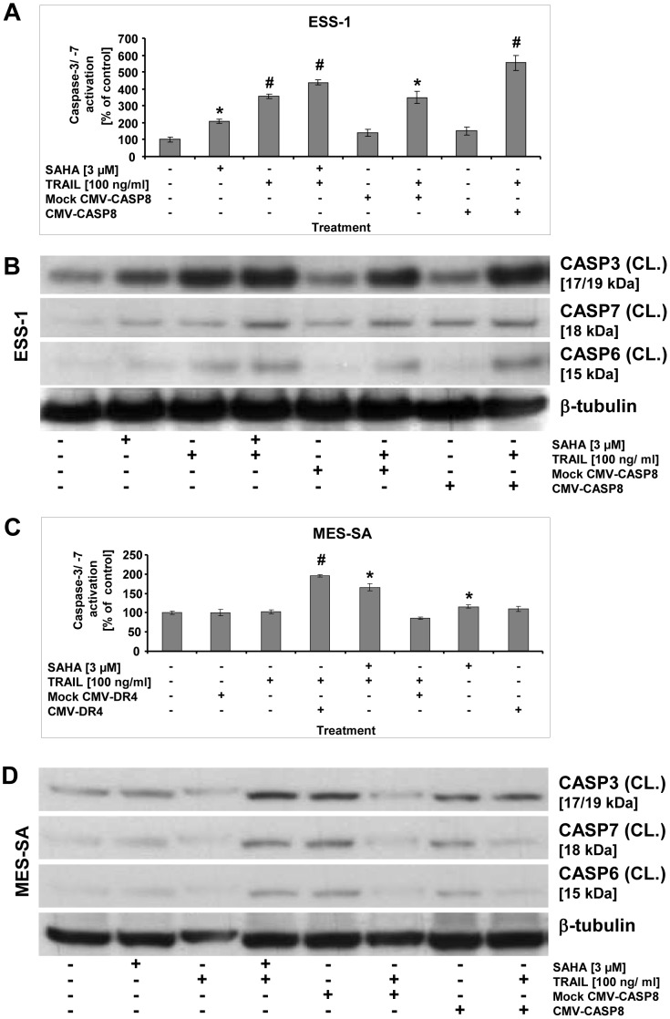
Figure 7. Reactivation of apoptosis by gene transfer in uterine sarcoma cells.
Measurements of apoptosis in uterine sarcoma cells by caspase-3/-7 activation that was reinduced by gene transfer (A and C). ESS-1 (A) and MES-SA cells (C) were transfected with caspase-8 (A) or DR4 (C) expression plasmids driven by a CMV promoter, respectively, and were supplemented with or without TRAIL before caspase-3/-7 activation was measured 24 hours later. Controls were mock-transfected and treated with or without TRAIL. For comparison, cells that received 3 μM SAHA and/or TRAIL were measured. Presented is the relative caspase activation in percentage as compared to untreated control cells. Asterisks (* p<0.05) or number signs (# p<0.001) indicate statistically significant differences as compared to the untreated control. Western blot analyses of activated executioner caspases of ESS-1 cells (B) and MES-SA cells (D) in order to observe apoptosis reinduction upon gene rescue experiments as demonstrated in (A). Samples with 50 μg (B) or 30 μg (D) protein were immunoblotted and analyzed with antibodies against cleaved (CL.) caspases-3, -6, -7, and β-tubulin as loading control. Untreated cells were used as control. The molecular weights of presented bands are indicated in brackets.

