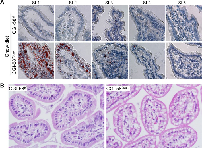
Figure 2. TG-rich LD accumulation in the cytoplasm of the proximal segment of small intestine of intestine-specific CGI-58 knockout mice.
The entire small intestine was collected and separated into 5 equal segments (SI-1 to SI-5, proximal to distal). A: Oil-red O staining of the 5 equal segments of small intestine from 6-month-old male CGI-58f/f and CGI-58f/f/cre mice on chow diet. B: Hematoxilin & eosin (H&E) staining of the first proximal segment of small intestine sections from CGI-58f/f and CGI-58f/f/cre mice on HFD.

