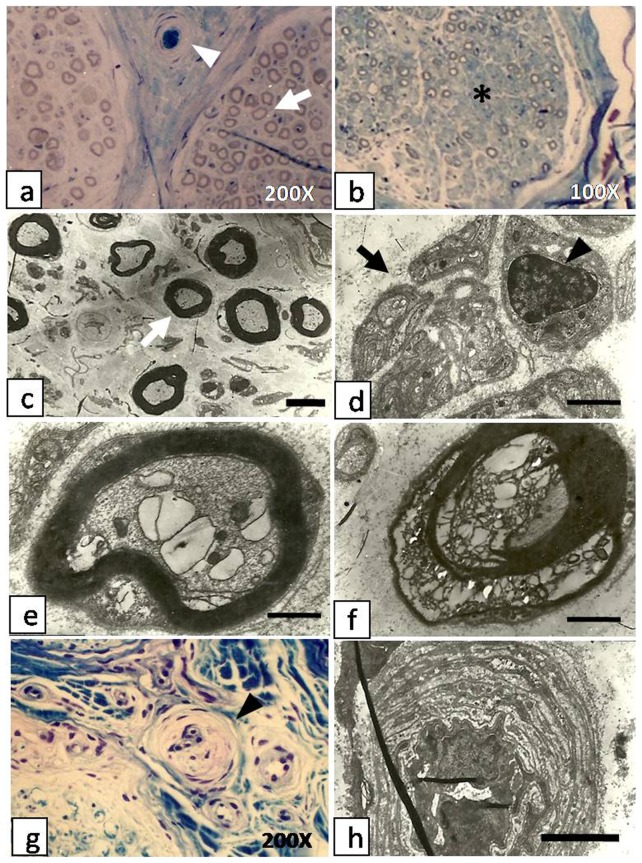
Figure 2. Cross section biopsies of sural nerves in patients with diabetic peripheral neuropathy.
a. Semi-thin section of sural nerve of case5 shows mild reduction in the number of nerve fibers (white arrow), and an arteriole plugged by a thrombus(white arrowhead). b. Semi-thin sections of sural nerve from case 1 shows severe loss of myelinated axons and increased endoneurial collagen (asterisk). C. and d. Semi-thin sections showdegeneration and regeneration of myelinated (white arrow) and unmyelinated (black arrow) fibers and proliferation of Schwann cells (black arrowhead) (case 3 and 6). e. and f. Semi-thin sections of a single myelinated axon showdegenerating myelin sheaths, absence of lamellar structures and balloon cell degeneration of the neuronal body (e: case 3); the axon shows almost complete structural disintegration (f: case 1). g. Section stained by Toluidine blue and examined by light microscopy shows luminal narrowing of epineural arterioles (black arrowhead). h. Electron micrograph of perineural arteriole shows luminal narrowing, endothelial thickening, basement membrane thickening and vascular wall thickening. Scale bar for (c–h): 2.5 μm, (d–f): 500 nm.

