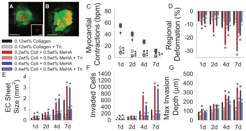
Fig. 4.
Pharmacological inhibition of myocardial contraction. (A–B) Live(green)-Dead(red) staining on explants and cells seeded on 0.12wt% collagen with either no treatment (A) or 1.5mM tricaine (B). Inset in (A) shows negative control, 4% PFA treatment for 5min. (C) Quantification of myocardial contractions with tricaine treatment. (D) Regional gel deformation induced by explants treated with tricaine. (E) EC sheet areas for tricaine treated explants. (F) EMT quantification via number of invaded cells in the presence of tricaine. (G) Maximum invasion depth of transformed cells. All data presented as average ± SEM and represents 3 technical replicates with n ≥ 6 biological replicates. *p < 0.05 vs. 0.12wt% collagen at same timepoint. ^p < 0.05 vs. same gel type at same time point.

