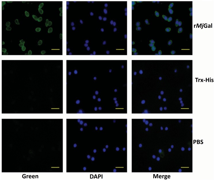
Figure 7. In vivo binding of rMjGal to shrimp hemocytes.
rMjGal, Trx-His, or PBS were injected into shrimp and the hemocytes were collected and analyzed by immunohistochemistry. The left panel indicated the binding signal detected by the anti-Histidine antibody for the recombinant protein (Green), the middle panel showed the hemocyte nucleus location (Blue), and the right panel showed the merge of previous two panels. Bar = 20 µm.

