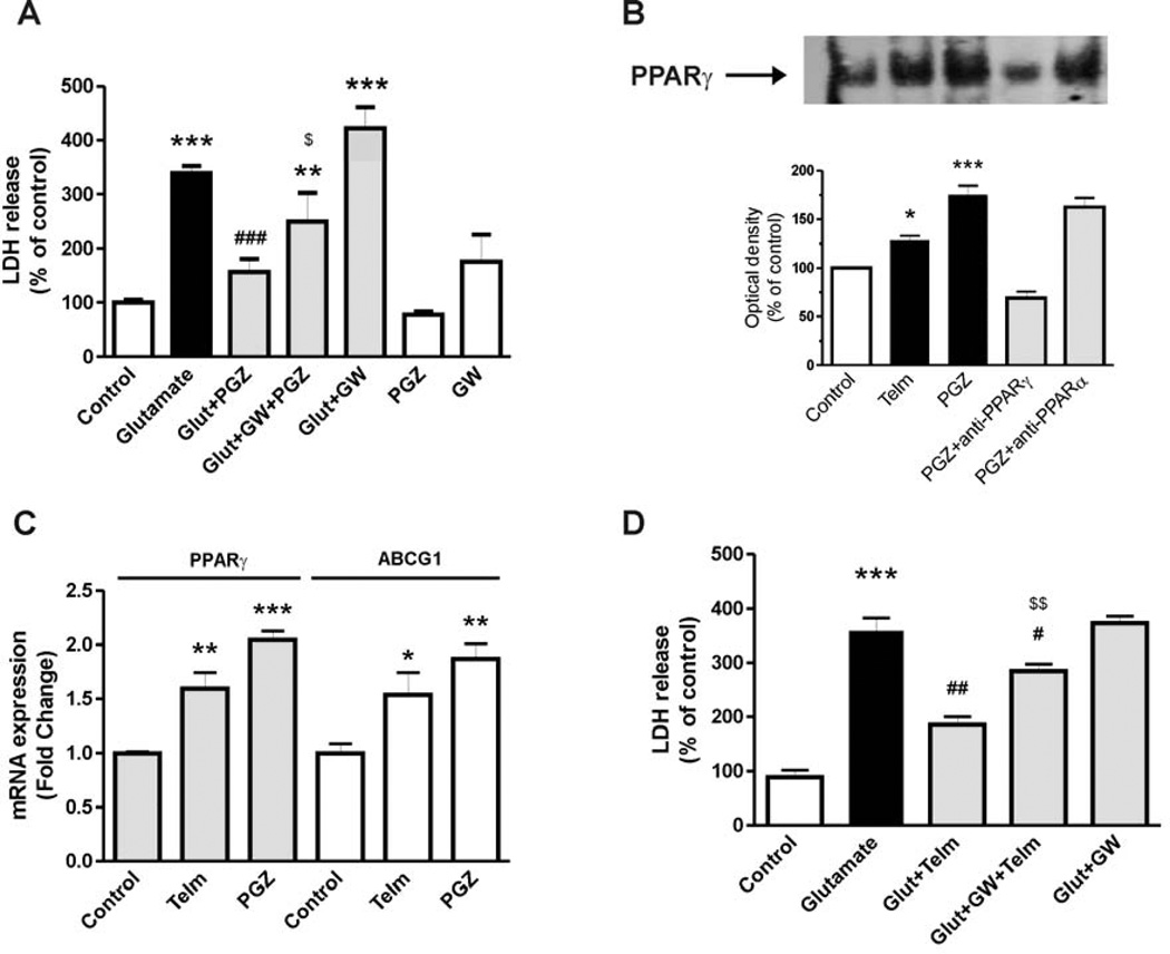
Fig. 6.
PPARγ activation is partially involved in the neuroprotective effect of telmisartan in rat CGCs. (A) CGCs were pre-treated with the PPARγ agonist pioglitazone (PGZ) (10 µM) for 2 hours followed by exposure to glutamate (Glut) for 24 hours. The PPARγ antagonist GW9662 (GW, 20 µM) was added 2 hours before PGZ treatment. (B) CGCs were treated with 1 µM telmisartan (Telm) or 10 µM pioglitazone (PGZ) for 4 hours to determine nuclear PPARγ activity using the Electrophoretic Mobility Shift Assay. Figure is a representative picture showing PPARγ-DNA binding. Intensity was measured by densitometry for quantitative analysis. Anti-PPARγ and anti-PPARα antibodies (2 µg) were used to determine the specificity of the shift. (C) CGCs were treated with 1 µM telmisartan or 10 µM pioglitazone for 24 hours to determine expression of the PPARγ target gene ABCG1. (D) CGCs were pre-treated with 20 µM GW9662 for 2 hours, followed by 2 hours exposure to telmisartan, followed by exposure to glutamate for 24 hours. Data are presented as means ±SEM of three independent experiments. *P < 0.05, ***P < 0.001 vs. Control; #P < 0.05, ##P < 0.01, ###P < 0.001 vs. Glutamate; $P < 0.05 vs. Glut+PGZ; $$P < 0.01 vs. Glut+Telm.

