Figure 3.
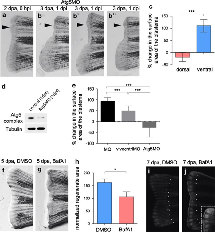
Macroautophagy is required for caudal fin regeneration. (a–c) When Atg5MO was injected into dorsal areas of 2-dpa blastemas, regeneration was severely compromised 1 day after the injection, compared with uninjected ventral fins (black arrowheads indicate site of injection). On panels b′ and b′′, additional examples of 3 dpa, 1 day post injection regenerates are shown. Statistical analysis (c) was performed for n=9 fins, P<0.001 (indicated by ***), paired t-test. (d) The efficacy of the Atg5MO was tested in western blots. In 1-dpf embryos the intensity of the band demarking the Atg12-Atg5 complex was reduced to less than 20%, indicating a potent inhibition of the Atg5 translation. (e) The effectiveness of the Atg5MO in inhibiting regeneration was significant compared with MilliQ water (MQ) and a standard control vivo MO (vivocntrlMO). (Statistical analysis was performed for n=5 fins from isogenic wild-type tue background fish for each treatment, P<0.001 for each pairwise comparison (indicated by ***), unpaired t-test.) (f–h) Regenerating fins were treated with either DMSO (f) or bafilomycin A1 (g), a potent inhibitor of autophagosome-autolysosome fusion. The latter treatment significantly impaired regeneration (h) at 5 dpa (n=6 fins, P<0.05, unpaired t-test). (i and j) When the fins of GFP-Lc3 transgenic fish were amputated, treatment with bafilomycin A1 led to a massive accumulation of the reporter in the blastema (i), as compared with DMSO controls (i). Inset in (j) shows the dorsal area of the representative fin at higher magnification. Dotted lines indicate amputation sites. Dpa, days post amputation; hpi, hours post injection; dpi, days post injection. Error bars refer to S.E.M.

