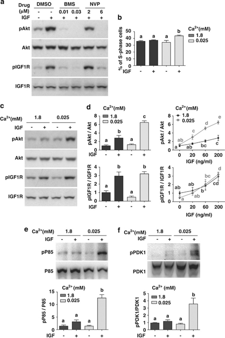
Figure 7.
Low extracellular [Ca2+] amplifies IGF signaling in human colon cells. (a) Caco-2 cells are responsive to IGF stimulation under low [Ca2+]. Confluent Caco-2 cells were incubated in serum-free medium containing 0.025 mM [Ca2+] overnight together with the indicated inhibitors at the indicated concentrations. IGF-2 (200 ng/ml) was added. Cells were lysed 10 min later and analyzed by western blot using the indicated antibodies. Similar results were found with IGF-1 (100 ng/ml). (b) Low extracellular [Ca2+] treatment increases the mitotic response to IGF stimulation. Confluent Caco-2 cells were incubated overnight in serum-free medium containing the indicated [Ca2+]. They were treated with 500 ng/ml IGF-1 and analyzed by FACS 96 h later. The percentage of S-phase was calculated and shown. Values shown are mean±S.D., n=3. Groups labeled with different letters are significantly different from each other (P<0.001). (c) Low [Ca2+] amplifies IGF-induced Akt signaling. Confluent Caco-2 cells were incubated in serum-free medium containing the indicated [Ca2+] overnight. IGF-2 (200 ng/ml) was added and cells were lysed 10 min later. The levels of pAkt, total Akt, pIGF1R, and total IGF1R were analyzed by western blot and their ratios calculated. Representative results are shown in the left panel and quantification results in the right panel. Values shown here and thereafter are mean±S.D., n=3–4. Groups sharing no common letters are significantly different from each other (P<0.05). (d) Dose-dependent effect of IGF-2 on Akt (upper panel) and IGF1R phosphorylation (lower panel). The cells were treated the same way as described in (c) with the indicated concentrations of IGF-2. (e) Low extracellular [Ca2+] amplifies IGF-induced PI3K signaling. The experiments were the same as (c). Cell lysates (1 mg) were precipitated with the 4G10 antibody followed by western blot analysis using a p85 antibody. Total p85 levels were determined by western blot. Representative results are shown in the upper panel. The ratio of phospho-p85 and total p85 was calculated and shown in the lower panel. Reciprocal co-IP experiments showed similar results (Supplementary Figure S7b). (f) Low extracellular [Ca2+] amplifies IGF-induced PDK1 signaling. Confluent Caco-2 cells were incubated in serum-free medium containing the indicated [Ca2+] overnight. IGF-1 (500 ng/ml) was added and cells were lysed 10 min later. The levels of phospho-PDK1 and total PDK1 were determined by co-IP experiments using the 4G10 antibody and a PDK1 antibody. Representative results are shown in the upper panel and quantitative results in the lower panel. IGF-2 treatment resulted in similar amplification of PDK1 signaling (Supplementary Figure S7c)

