Abstract
Adipocyte cell number is a crucial factor for controlling of body weight and metabolic function. The regulation of adipocyte numbers in the adult organism is not fully understood but is considered to depend on the homeostasis of cell differentiation and apoptosis. Herein, we show that targeted deletion of the activator protein (AP-1)-related transcription factor Fra-2 in adipocytes in vivo (Fra-2Δadip mice) induces a high-turnover phenotype with increased differentiation and apoptosis of adipocytes, leading to a decrease in body weight and fat pad mass. Importantly, adipocyte cell numbers were significantly reduced in Fra-2Δadip mice. At the molecular level, Fra-2 directly binds to the PPARγ2 promoter and represses PPARγ2 expression. Deletion of Fra-2 leads to increased PPARγ2 expression and adipocyte differentiation as well as increased adipocyte apoptosis through upregulation of hypoxia-inducible factors (HIFs). These findings suggest that Fra-2 is an important checkpoint to control adipocyte turnover. Therefore, inhibition of Fra-2 may emerge as a useful strategy to increase adipocyte turnover and to reduce adipocyte numbers and fat mass in the body.
Keywords: AP-1/Fra-2, HIFs, adipocyte, apoptosis
Alterations in adipose tissue lead to severe metabolic disturbances. During obesity, white adipose tissue expansion is based on both cellular hypertrophy (an increase in adipocyte volume) and hyperplasia (an increase in adipocyte cell number). White adipose tissue becomes hypoxic during obesity that leads to adipose tissue dysfunction.1 The response of most cells to hypoxia is to induce a program of gene expression that is regulated by hypoxia-inducible factors (HIFs), basic helix-loop-helix/PAS transcription factors consisting of a α-subunit (HIF1α) and a β-subunit (HIF1β).2 HIF target genes are involved in several physiological processes such as angiogenesis, glucose metabolism and cell survival.3
In accordance, targeted deletion of HIF1α in adipocytes has an impact on dietary obesity and associated pathologies such as glucose intolerance.4, 5 However, the role of HIFs during adipocyte differentiation and survival remains defined incompletely to date. Moreover, despite that adipocyte hypertrophy prevails in obesity, the adipocyte number variation in adult age remains under debate.1 Apoptosis is a normal phenomenon of cell death for the purpose of maintaining homeostasis. The apoptotic molecular machinery such as the mitochondrial and the caspase pathways are present in adipocyte and not different from other cells. It is reported that several adipokines play roles in induction of adipocyte apoptosis.6 However, the question remains of how apoptosis is regulated in adipocytes at the cellular and molecular levels.
Mesenchymal stem cells (MSCs) can differentiate into adipocytes following the induction of lineage-specific transcription factors C/EBPβ (CCAAT/enhancer binding protein-β) that induce the expression of PPARγ2 (peroxisome proliferator-activated receptor-γ2) and C/EBPα controlling the late stages of adipogenesis.7 Another transcription factor family that can influence adipocyte commitment is the activator protein-1 (AP-1) transcription factor complex. This complex consists of a variety of dimers composed of members of the Fos, Jun and activating transcription factor (ATF) families.8 Analyses of mice genetically modified for Fos proteins have underlined the important functions of Fos in osteoblast and adipocyte differentiation.8 Mice overexpressing Fos-related antigen 1 (Fra-1) or the short isoform of FosB (ΔFosB) show impaired adipocyte differentiation9, 10, 11 but enhanced osteoblast differentiation.12, 13, 14
Interestingly, the second Fra family member, Fos-related antigen 2 (Fra-2), regulates bone mass via affecting HIF expression. Thus, mice overexpressing Fra-2 develop an osteosclerotic phenotype, whereas the absence of Fra-2 leads to a defect of osteoblast differentiation15 and a giant osteoclast phenotype induced by the activation of HIF1α.16 Furthermore, leptin is a transcriptional target of Fra-2,17 suggesting that Fra-2 may indeed represent an important transcription factor for adipocytes. Herein, we show that Fra-2 deletion in adipocytes affects body and fat pad mass by the regulation of adipocyte number. Mechanistically, Fra-2 regulates transcriptionally PPARγ2 expression and adipocyte survival by the modulation of HIF expression.
Results
Inducible adipocyte-specific deletion of Fra-2
To obtain functional data for a role of Fra-2 in the adipose tissue, we generated adipocyte-specific Fra-2 knockout mice. Mice with the Fabp4-CreERT allele were crossed with mice carrying Fra-2 floxed alleles to delete Fra-2 in adipocytes (Fra-2Δadip). As Fabp4-CreERT is a tamoxifen-inducible line, we were able to analyze the role of Fra-2 inactivation in adult mice. Injection of tamoxifen was performed at the age of 6 weeks and the analyses were performed 6 weeks later. Immunohistochemical (IHC) staining for Fra-2 of fat tissues showed decreased Fra-2 expression in adipocytes of white and brown adipose tissue, whereas no decrease in expression was found in the spleen (Supplementary Figure 1A). Furthermore, only very low levels of Fra-2 mRNA were expressed in the white and brown adipose tissue of Fra-2Δadip mice (Supplementary Figure 1B), whereas its expression in liver, muscle, spleen and lung was equal to wild-type littermate controls (Supplementary Figure 1B). These results indicate that Fra-2 deletion in Fra-2Δadip mice is specific to the adipose tissue.
Fra-2 deletion in adipocytes decreases body and fat pad weight
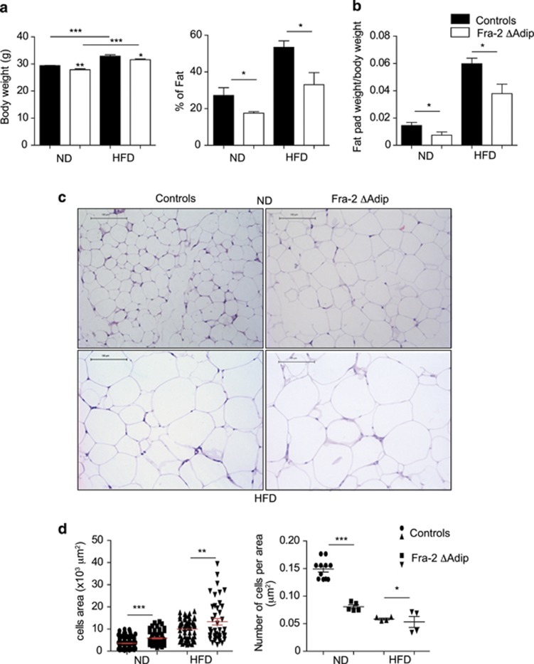
We first assessed body and fat pad weight in Fra-2 wild-type and mutant mice. Total body weight and percentage of body fat were significantly decreased in Fra-2Δadip mice compared with wild-type controls in conditions of normal diet (ND) (Figure 1a). Similarly, the ratio of fat pad weight to body weight was significantly lower in Fra-2Δadip mice than in controls (Figure 1b). When mice were fed a high-fat diet (HFD) for a period of 6 weeks for inducing obesity, similar changes were observed: Fra-2Δadip mice showed significantly lower body weight and fat pad weight than wild-type controls (Figures 1a and b).
Figure 1.
Fra-2 Δadip mice have decreased body weight and adipocyte number. (a) Body weight of mice and fat content from controls and Fra-2 Δadip mice at 6 weeks after tamoxifen injections with mice receiving normal diet (ND) or high-fat diet (HFD) (n=4–5). (b) Fat pad per body weight of Fra-2Δadip mice and littermate controls at 6 weeks after tamoxifen injections with mice receiving ND or HFD (n=4–5). (c) Hematoxylin and eosin staining of fat pad tissue from Fra-2Δadip mice and littermate controls at 6 weeks after tamoxifen injection receiving ND or HFD. Magnification × 20, bars=100 μm. (d) Adipocyte cells area and number of adipocyte per area of Fra-2Δadip mice and littermate controls at 6 weeks after tamoxifen injections with mice under ND or HFD (n=4–10). Bars represent mean values±S.D. Statistical analyses: *P<0.05, **P<0.01, ***P<0.001
Low adipocyte number but increased adipocyte size in Fra-2 Δadip mice
Further histological analyses of the fat pads of Fra-2Δadip mice and respective controls showed that white adipocyte cell number was significantly decreased in Fra-2Δadip mice compared with controls when mice received ND (Figures 1c and d). Furthermore, analysis of adipocyte cell size showed a significant increase in cell size despite smaller cell number in Fra-2Δadip compared with controls (Figures 1c and d). HFD increased adipocyte size in both mutant and wild-type mice, reflecting hypertrophy of adipocytes that was significantly more pronounced in Fra-2 mutants than controls (Figures 1c and d).
Increased adipocyte apoptosis in Fra-2Δadip mice
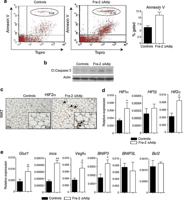
To better understand the molecular mechanisms behind the fat phenotype observed in Fra-2Δadip mice and to determine why adipocyte numbers are low in Fra-2 mutants, we analyzed the proliferation and the apoptosis status of the fat pad. No differences could be detected in proliferation of adipocyte as assessed by Ki67 staining (data not shown). However, apoptosis assessed by TUNEL assay, annexin V flow cytometry, cleaved caspase 3 staining and western blot revealed an increased rate of apoptosis in the fat pad of Fra-2Δadip mice (Figures 2a and b and Supplementary Figure 2A), suggesting that low fat pad mass and low adipocyte numbers in Fra-2Δadip mice depend on increased adipocyte apoptosis.
Figure 2.
Fra-2Δadip fat pads are hypoxic and show higher apoptosis rates. (a) Quantification of apoptotic cells by FACS in perigonadal fat from Fra-2Δadip and littermate control mice at 6 weeks after tamoxifen injection (n=4). (b) Western blot for cleaved caspase-3 in perigonadal fat from Fra-2Δadip and littermate control mice at 6 weeks after tamoxifen injection. (c) HIF2α staining in Fra-2Δadip and littermate controls fat pad at 6 weeks after tamoxifen injection. Magnification × 20, insert × 40. Black arrows indicate HIF-positive cells. (d) Real-time PCR analyses of HIF1α, HIF1β and HIF2α in Fra-2Δadip and littermate controls fat pad at 6 weeks after tamoxifen injection (n=4). (e) Real-time PCR analyses of HIFs target genes Glut1, Inos, Vegfα, BNIP3, BNIP3L and Bcl2 in Fra-2Δadip and littermate controls fat pad at 6 weeks after tamoxifen injection (n=4). Bars represent mean values±S.D. Statistical analyses: *P<0.05, **P<0.01
Increased hypoxia and HIF activation in adipose tissue of Fra-2Δadip mice
Our previous finding showing that Fra-2 is involved in the regulation of osteoclast apoptosis and that this effect depends on HIF1α activation16 prompted us to investigate whether the increased apoptosis in Fra-2Δadip mice was based on HIF activation and tissue hypoxia. Indeed, in vivo analysis revealed increased hypoxia in the fat pad tissue of Fra-2Δadip mice compared with controls (Supplementary Figure 2B). IHC stainings of fat pad tissue showed an increased number of HIF1α- and HIF2α-positive cells in Fra-2Δadip mice compared with controls (Supplementary Figure 2B and Figure 2c). In addition, the molecular analyses of HIFs by quantitative PCR (qPCR) revealed increased expression of HIF1α and HIF2α in Fra-2Δadip fat pad, whereas no differences in HIF1β levels were detected (Figure 2d). Moreover, expression of several HIF target genes such as Glut1 (glucose transporter), Inos (nitric oxide synthase), Vegfα (vascular endothelial growth factor α) and BNIP3 (BCL2/adenovirus E1B 19 kDa protein-interacting protein 3) was increased in Fra-2Δadip fat pads, whereas others such as Bcl2 (B-cell lymphoma 2) and BNIP3L (BCL2/adenovirus E1B 19 kDa protein-interacting protein 3-like) were not altered (Figure 2e). These findings support the concept that Fra-2 regulates hypoxia and HIF expression in the adipose tissue that affects adipocyte numbers and fat pad size via regulation of apoptosis.
Hypoxia drives expression of HIFs and apoptosis-related genes in adipocytes
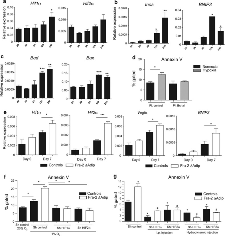
In order to determine whether hypoxia could induce apoptosis in a cell autonomous manner, adipocytes isolated from fat pads were placed in hypoxic chambers and analyzed for HIFs and HIF target gene expression. In addition, expression of apoptosis-related genes was analyzed. As shown in Figures 3a and b, HIF1α, HIF2α and their target genes Inos and BNIP3 were increased in adipocytes placed in hypoxic chambers for 24 h. Moreover, Bad (Bcl-2-associated death promoter) and Bax (Bcl-2-associated X protein) mRNA levels in adipocytes increased within 12 h of hypoxia (Figure 3c), suggesting that hypoxic conditions lead to an imbalance of the Bcl-2 family in favor of the apoptotic pathway.
Figure 3.
Hypoxia drives adipocyte apoptosis and Fra-2 regulates HIF expression in cell autonomous manner. (a and b) Real-time PCR analyses of HIF1α and HIF2α (a) and HIF targets Inos and BNIP3 (b) in primary adipocytes placed in hypoxic chambers for 0, 3, 6, 12 and 24 h (n=3). (c) Real-time PCR analyses of Bad and Bax in primary adipocytes placed in hypoxic chambers for 0, 3, 6, 12 and 24 h (n=3). (d) Quantification of apoptotic cells by FACS in primary adipocytes transfected with control or overexpressing Bcl-xl plasmid under normoxia or hypoxia for 24 h (n=3). (e) Real-time PCR analyses of HIF1α, HIF2α, Vegfα and BNIP3 in adipocytes of Fra-2Δadip and controls cultures at days 0 and 7 of in vitro differentiation (n=3). Bars represent mean values±S.D. Statistical analyses: *P<0.05, **P<0.01,***P<0.001. (f) Quantification of apoptotic cells by FACS in primary adipocytes isolated from Fra-2Δadip and controls fat pad and transfected with Sh control or Sh plasmid against HIF1α or HIF2α under hypoxia for 24 h (n=3). (g) Quantification of apoptotic cells by FACS in perigonadal tissue isolated from Fra-2Δadip and control mice after i.p. or hydrodynamic injection with Sh control or Sh plasmid against HIF1α or HIF2α (n=3). *P-values compared with controls. #P-values compared with Fra-2Δadip adipocytes. Statistical analyses: * or #P<0.05, **P<0.01, ***P<0.001
To determine whether apoptosis was dependent on the Bcl-2 family, overexpressing plasmids for B-cell lymphoma-extra large (Bcl-xl) were transfected in primary adipocyte exposed to normoxia or hypoxia 1% for 24 h. The annexin V flow cytometry analyses revealed increased apoptosis in adipocyte culture under hypoxia that can be rescued by the overexpression of Bcl-xl (Figure 3d), suggesting that the apoptosis induced by hypoxia in adipocytes is dependent on the Bcl-2 family.
Fra-2 regulates HIFs in the adipocyte in a cell autonomous manner
In order to determine whether Fra-2 affects HIF expression in adipocytes, we analyzed HIF expression and HIF target gene expression on adipocyte-derived stem cells isolated from fat pads of Fra-2Δadip and wild-type controls. Interestingly, HIF1α, HIF2α and their targets Vegfα and BNIP3 were significantly increased in the Fra-2Δadip cultures at day 7 of differentiation (Figure 3e). These data suggest that Fra-2 deletion leads to increased HIFs and HIF target gene expression in the adipose tissue in vivo and in vitro.
In order to determine whether apoptosis induced by hypoxia was dependent on HIF1α or HIF2α or both, knockdown of HIF1α and HIF2α was performed in vitro by transfection of primary cells isolated from Fra-2Δadip and control fat pads. Interestingly, the knockdown of HIF1α or HIF2α was able to rescue the increased level of apoptosis observed in Fra-2Δadip adipocyte cultures under hypoxic conditions (Figure 3f). Moreover, to determine whether similar observations could be found in vivo, i.p. and hydrodynamic injections of shRNA against HIF1α or HIF 2α were performed in Fra-2Δadip and littermate control mice. Interestingly, in wild-type condition, the knockdown of HIF1α and HIF2α was able to decrease the basal level of apoptosis in perigonadal fat tissue (Figure 3g). In addition, no difference in apoptosis could be detected anymore in Fra-2Δadip and littermate control perigonadal fat after knockdown of HIF1α or HIF2α (Figure 3g), suggesting that HIF1α and HIF2α regulate apoptosis in white adipose tissue.
Increased PPARγ2 expression as the link between Fra-2 and HIF
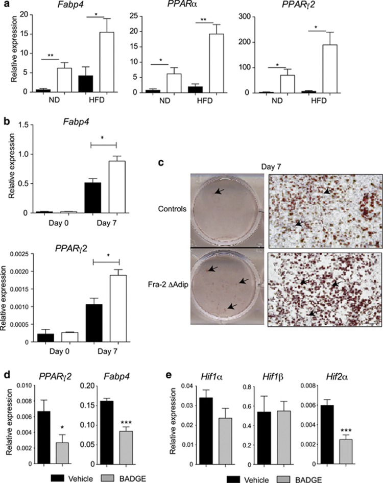
To explain the molecular mechanisms of how Fra-2 regulates HIFs, we checked for differences in the regulation of genes involved in adipocyte differentiation among Fra-2Δadip mice and controls. In vivo, no significant changes could be detected in the expression of C/ebpβ, C/ebpα, C/ebpδ, Glut4, PPARα, PPARγ1, adiponectin and resistin (Supplementary Figure 2C and D). Interestingly, increase in mRNA levels of PPARγ total and PPARγ2 as well as Fabp4 (fatty acid binding protein 4) was observed in the fat pad from Fra-2Δadip mice fed ND and HFD as compared with respective wild-type littermate controls (Figure 4a and Supplementary Figure 2C). IHC analysis for PPARγ expression confirmed its upregulation at the protein levels (Supplementary Figure 2E).
Figure 4.
PPARγ2 is regulated by Fra-2 and can regulate HIF expression. (a) Real-time PCR analyses of Fabp4, PPARα and PPARγ2 in fat pad tissue from Fra-2Δadip mice and littermate controls at 6 weeks after tamoxifen injections with mice receiving normal diet (ND) or high-fat diet (HFD) (n=4–5). (b) Real-time PCR analyses of Fabp4 and PPARγ2 in adipocytes of Fra-2Δadip and controls cultures at days 0 and 7 of in vitro differentiation (n=3). (c) Oil Red O staining of adipocytes derived from fat pad tissue of Fra-2Δadip mice and littermate controls at day 7 of in vitro differentiation. Bars represent 200 μm. Arrows indicate adipocytes. (d and e) Real-time PCR analyses of PPARγ2, Fabp4 (d) and HIF1α, HIF1β and HIF2α (e) in primary adipocytes treated with vehicle or BADGE (n=3). Bars represent mean values±S.D. Statistical analyses: *P<0.05, **P<0.01, ***P<0.001
To determine whether the same phenotype could be observed in another white adipose tissue, subcutaneous fat from Fra-2Δadip and wild-type littermate control mice was analyzed. Indeed, similar phenotype, with increased adipocyte size, increased apoptosis, increased hypoxic markers and increased Fabp4 and PPARγ2 mRNA levels, was observed in the subcutaneous white fat from Fra-2Δadip mice when compared with littermate controls (Supplementary Figure 3), suggesting a common pathway in different white fat tissues.
Next, adipocyte differentiation and markers were analyzed when adipocyte stem cells from Fra-2Δadip mice and respective controls were isolated from the fat pad and differentiated in vitro. Whereas C/ebpα, C/ebpα C/ebpδ and PPARγ1 were not different among cells from mutant and control mice, the mRNA expression of Fra-2 was decreased and mRNA expression of adiponectin, Fabp4, total PPARγ, and PPARγ2 was significantly increased in Fra-2Δadip cells at day 7 of differentiation (Supplementary Figure 4 and Figure 4b). In addition, cell differentiation was significantly increased in Fra-2Δadip cultures compared with controls, potentially explaining the increased adipocyte size observed in Fra-2Δadip mice (Figure 4c).
To determine whether PPARγ2 can regulate HIFs in adipocytes, inhibition of PPARγ2 activity by BADGE was performed (Figure 4d). Treatment with BADGE not only led to a decrease of the adipocyte marker Fabp4 (Figure 4d), but also to significant suppression of HIF2α expression, whereas the expression of HIF1α remained unchanged (Figure 4e).
Fra-2 leads to direct transcriptional repression of PPARγ2
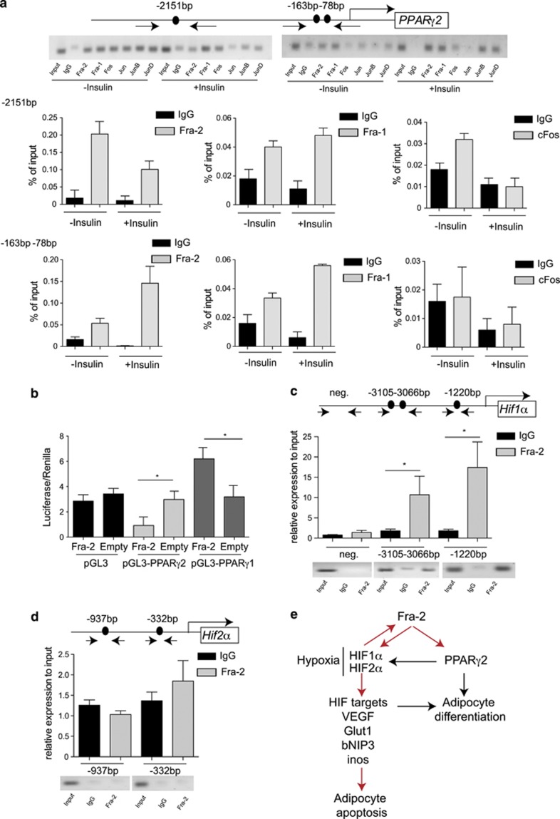
As PPARγ2 essentially controls adipocyte differentiation as well as HIF activation in adipocytes,18 we reasoned that PPARγ2 was the key gene regulated by Fra-2 in adipocytes. We therefore assessed Fra-2 binding to the PPARγ2 promoter by chromatin immunoprecipitation (ChIP) analysis in 3T3L1 cell lines treated with insulin for 24 h. As shown in Figure 5a, specific primer pairs were used to amplify DNA fragments containing the AP-1 consensus TRE element. Fos and Jun binding to the PPARγ2 promoter was observed in two potential sites (Figure 5a and Supplementary Figure 5A). Interestingly, at the sites −163/−78 bp, the binding of Fra-2 and Fra-1 is increased after insulin treatment (Figure 5a), suggesting that Fra-2 can bind the PPARγ2 promoter and regulate its expression following insulin treatment.
Figure 5.
PPARγ2 and HIF1α are transcriptional targets of Fra-2. (a) Chromatin immunoprecipitation (ChIP) for PPARγ2 promoter. Arrows indicate primers amplifying fragments for the TRE elements. Chromatin isolated from 3T3L1 cells with insulin treatment for 24 h was immunoprecipitated with IgG and AP-1 antibodies. Real-time PCR analyses and loading of the gel are shown (n=2). (b) Luciferase assay analyses of Fra-2 on PPARγ1 and PPARγ2 promoter; empty plasmids are shown for the control experiment. (c and d) ChIP for HIF1α (c) and HIF2α (d) promoters. Arrows indicate primers amplifying fragments for the TRE elements. Chromatin from 3T3L1 cells was immunoprecipitated with IgG and Fra-2 antibodies. Real-time PCR analyses and loading of the gel are shown (n=3). (e) Scheme of Fra-2 actions in adipocytes. Red arrows indicate findings from the manuscript and black arrows indicate knowledge from the literature. Bars represent mean values±S.D. Statistical analyses: *P<0.05
Moreover, in DNA co-transfection experiments, Fra-2 expression vector decreased the activity of a luciferase reporter containing the human PPARγ2 promoter fragment including the two sites −163/−78 bp (Figure 5b). In contrast, an increase in the luciferase activity could be detected when DNA co-transfection experiments were performed with PPARγ1 promoter fragment (Figure 5b). These findings, together with the altered expression of PPARγ2 in Fra-2Δadip mutants, indicate that PPARγ2 is a direct transcriptional target of Fra-2.
Fra-2 and PPARγ2 can regulate HIFs and vice versa
In order to determine whether HIF1α and HIF2α could be transcriptionally regulated by Fra-2, we performed ChIP analysis in 3T3L1, on AP-1 consensus elements found in both promoters. Interestingly we could find that Fra-2 was able to bind the HIF1α promoter at two putative TRE sequences, −3105/−3066 and −1220 bp, before the starting codon (Figure 5c), whereas no binding of Fra-2 could be detected on the HIF2α promoter (Figure 5d), suggesting a direct transcriptional activation of HIF1α by Fra-2.
To understand how HIF2α can be regulated, we performed the ChIP for PPARγ on the previously analyzed sequences. No PPARγ binding could be detected on HIF1α promoter (Supplementary Figure 5B). Surprisingly PPARγ could bind the HIF2α promoter at the site −937 bp (Supplementary Figure 5C). These results suggest that Fra-2 and PPARγ can regulate HIF1α and HIF2α respectively.
To determine whether HIFs can regulate Fra-2 as a retro-control mechanism, its mRNA levels were analyzed in 3T3L1 cells exposed to hypoxia. Interestingly, Fra-2 mRNA level was upregulated in 3T3L1 cell culture in hypoxic conditions (Supplementary Figure 6), suggesting a compensatory mechanism on Fra-2 induced by hypoxia in adipocytes.
Discussion
The results presented in this study reveal a novel function of Fra-2/AP-1 in adipocytes, that is, to regulate fat pad mass and adipocyte number. Mechanistically, Fra-2 inhibits the expression of PPARγ2, a key regulator of adipocyte differentiation and homeostasis. By repressing PPARγ2, Fra-2 also represses HIFs that control hypoxia and adipocyte survival (see scheme in Figure 5e). The control of adipocyte numbers has been a matter of uncertainty since many years.1 Here we found a new pathway that is able to determine adipocyte number in adult age by the regulation of Fra-2 and HIFs.
It can be hypothesized that the regulation of the balance between adipocyte differentiation and death determines adipocyte numbers in the fat tissue. Differentiation of mesenchymal cells into adipocytes involves a cascade of transcription factors such as C/EBPα and C/EBPα that in turn activate PPARγ, an essential gene for the activation of several adipocyte-specific genes.19 In addition, AP-1 proteins, such as Fra-1 and ΔFosB, have also been shown to regulate adipogenesis through C/EBPα or C/EBPβ expression respectively, thereby decreasing adipocyte differentiation.10, 11 Moreover, Fra-2 was shown to regulate leptin expression in adipocytes and osteocalcin in osteoblasts, both of which are involved in the control of body weight and fat distribution.15, 17 By using Fabp4-CreERT tamoxifen-inducible line, we were able to analyze the role of Fra-2 inactivation in adult mice. The Fabp4-Cre lines were known to potentially have recombination in nonendothelial and nonmyocyte cells in the skeletal muscle.20 Despite no difference in Fra-2 levels in other organs in the mutant mice, we could not exclude indirect effects induced by unwanted deletion. However, our biochemistry and in vitro culture studies directly link Fra-2 expression to adipogenesis by the transcriptional inhibition of PPARγ2 expression.
PPARγ2 is not only a master regulator of adipogenesis, but can also display a role in apoptotic signal transduction in adipocytes.6 Adipocytes express several molecules of the intrinsic and extrinsic apoptotic pathways. An excess of apoptosis can lead to lipodystrophic metabolic changes.21 However, induction of moderate amounts of adipocyte apoptosis in obese mice has shown metabolic improvement,22 suggesting that modulation of fat homeostasis by regulating differentiation and apoptosis might represent an instrument for controlling fat pad mass in the body. Interestingly, PPARγ can have proapoptotic functions in adipocytes. Thus, PPARγ is directly involved in the leptin-induced adipocyte apoptosis signal pathway.23 Our observation that increased PPARγ expression in the absence of Fra-2 not only enhanced adipocyte differentiation but also apoptosis support this concept.
During hypoxia and induction of HIF1α, an intricate balance exists between factors that induce or counteract apoptosis. Previous studies of gain and loss of function of HIF1α in adipocytes have revealed its role during adipocyte differentiation and diabetes.24, 25 In addition, HIF2α, which has independent functions of HIF1α, increases during adipogenesis in vitro, indicating that its upregulation is necessary for execution of adipogenesis and maintenance of mature adipocyte functions.26 Our results confirmed that increased HIF1α and HIF2α expression correlated with a cell-autonomous increase of adipocyte differentiation. In addition, we show that hypoxia and increased HIF expression induced adipocyte apoptosis that was dependent on Bcl-xl. Notably, HIF1α can directly be regulated by Fra-2 and HIF2α expression that, in turn, is directly regulated by PPARγ, providing a link between adipocyte differentiation and apoptosis. Moreover, the alteration of Fra-2 expression by hypoxia as a feedback loop provides further insight into the regulation of adipocyte turnover.
In summary, our data add a new role of Fra-2 in adipose tissue homeostasis by regulating PPARγ2 and HIF expression. Modulation of Fra-2 expression in mesenchymal cells may emerge as potent tool to control adipocyte number in the body and may open new avenues to manage obesity and metabolic disease.
Materials and Methods
Mice
The generation of Fra-2 floxed mice is described elsewhere.27, 28 Fabp4-CreERT mice were described previously.29 All mice were maintained on a mixed 129/C57Bl6 background. Littermate mice were used as controls in this study. Genotyping primers were as follows: Fra-2 lox forward: 5′-GAGGGAGTTGGGGATAGAGTGGTA-3′, Fra-2 lox reverse: 5′-GGACAGCAGGTCAGGAGTAGATGA-3′, Cre forward: 5′-CCAGAGACGGAAATCCATCGCTCG-3′ and Cre reverse: 5′-CGGTCGATGCAACGAGTGATGAGG-3′. To delete Fra-2 in vivo, mutant and control mice were injected intraperitoneally with 1 mg tamoxifen (Sigma-Aldrich Chemie GmbH, München, Germany) for 5 consecutive days.
Intraperitoneal and hydrodynamic injection of shRNA HIFs
To downregulate the expression of HIF1α and HIF2 α in vivo, intraperitoneal and hydrodynamic intravenous injections were performed with 200 μl or 1 ml respectively with shRNA-HIF1 or shRNA-HIF2. After 24 h for the i.p. experiment and 72 h for the hydrodynamic experiment, mice were killed and fat pad adipocytes apoptosis was analyzed.
Histological analysis and immunohistochemistry
Mice were killed by CO2 asphyxia and tissues were fixed in 3.7% PBS-buffered formaldehyde. For histological analysis, tissues were embedded in paraffin. Then, 5 μm sections of white adipose tissue and 1 μm sections of brown adipose tissue and spleen were used for histological analyses.
Immunohistochemical stainings for Fra-2, HIF1α, HIF2α (Abcam plc, Cambridge, UK), cleaved Caspase 3 (Cell Signaling Technology, Inc., Danvers, MA, USA) and PPARγ (Santa Cruz Biotechnology, Inc., Dallas, TX, USA) were performed on deparaffinized sections using Proteinase K as unmasking solution and according to the manufacturer's instructions for the Vectastain KIT (Vector Laboratories, Inc., Burlingame, CA, USA). TUNEL assay was performed according to the manufacturer's instructions using the In Situ cell death Detection kit (Roche Diagnostics Deutschland GmbH, Mannheim, Germany) . Fluorescein is detected by anti-fluorescein antibody Fab fragments from sheep, conjugated with horseradish peroxidase (POD) (Roche Diagnostics Deutschland GmbH) and detected by DAB (Dako, Hamburg, Germany).
Apoptosis analyses by flow cytometry
Apoptosis and necrosis were quantified in adipocytes isolated from fat pad tissue, by Annexin V-FITC (Invitrogen, Life Technologies GmbH, Darmstadt, Germany) staining in the presence of TO-PRO-3 Iodide (Invitrogen). Adipocytes were incubated for 20 min at room temperature with 200 μl Annexin V-FITC in Annexin-binding buffer, washed and resuspended in 200 μl Annexin-binding buffer together with TO-PRO-3 Iodide and measured by flow cytometry.
Hypoxia analysis
Hypoxia stainings were performed using the hypoxyKit (Chemicon, Millipore, Billerica, MA, USA) according to the manufacturer's instructions.
RNA isolation and reverse transcription
Total RNA was isolated with the TRIzol or TriFast protocol (Invitrogen and PEQLAB Biotechnologie GMBH, Erlangen, Germany, respectively). cDNA synthesis was performed using 0.50–2 μg RNA with the cDNA Reagents from Applied Biosystems (Life Technologies GmbH).
Real-time PCR analyses
The qPCR reactions were performed using SYBR Green (Eurogentec Deutschland GmbH, Köln, Germany). Primers for genes analyses and primers for ChIP analyses used for real-time PCR are available upon request. The comparative CT method was used to quantify the amplified fragments. RNA expression levels were analyzed in duplicate and normalized to actin as housekeeping gene, whereas chromatin-bound fragments were normalized to the same fragments amplified from input chromatin.
Cell culture
The adipogenic cells were generated by culturing adipocyte stem cells isolated from fat pad tissue. Fra-2 deletion in vitro was induced by treating the cells with 3 μg/ml tamoxifen for 2 days. Adipocyte differentiation was induced by addition of 5 μg/ml insulin, 1 μM dexamethasone and 500 μM 3-isobutyl-1-methylxanthine (IBMX) to the confluent cultures.
Adipocyte stem cells isolated from fat pad tissue were cultured in 12-well plates until 70–80% of density. Cells where transfected with 1 μg of shRNA HIF1α, shRNA HIF2α or overexpressing Bcl-xl plasmid diluted in the appropriate amount of Lipofectamine Reagent (Invitrogen). The transfected cells were incubated at 37 °C and 5% of CO2. After 48 h, cells were placed in hypoxic chambers (1% O2) for 24 h.
In ‘hypoxic' experiments, cells were placed in hypoxic chambers (1% O2) for 24 h.
In experiments using BADGE (Sigma), inhibitor of PPARγ, the cells were treated with the adipogenic cocktail containing 100 μM BAGDE for 7 days. Medium was changed daily. Isolation of RNA and staining with Oil Red O was performed on day 0 (confluency) and day 7 after stimulation.
Oil Red O staining
Cells were fixed in 3.7% PBS-buffered formaldehyde for 1 h. Cells were stained with Oil Red O solution (0.3% Oil Red O in 60% isopropanol) for 2 h.
Chromatin immunoprecipitation
PPARγ and HIF promoter analyses were performed by ‘Tfsearch' web site (http://www.cbrc.jp/research/db/TFSEARCH.html).
For ChIP analyses, 3T3L1 cells were stimulated or not with 5 μg/ml insulin for 24 h. ChIP was performed using the ChIP-IT Express kit (Active Motif, La Hulpe, Belgium) with antibodies against Fra-2, Fra-1, cFos, JunB, JunD (Santa Cruz Biotechnology, Inc.), cJun (Cell Signaling Technology, Inc.) and PPARγ (Santa Cruz Biotechnology, Inc.), and control experiments were performed with IgG antibody. Bound fragments were quantified by real-time PCR.
Reporter assay
The promoter-reporter vector for human PPARγ1 and 2 were provided by J Auwerx (Ecole Polytechnique Fédérale in Lausanne, Switzerland). Control experiments were performed with a pGL3 empty plasmid. 6 × 10⁴ Hek cells/well were plated in 24-well dishes. Then, 1 μg of the luciferase reporter construct, 0.2 μg of the Renilla internal control (phRG-TK, Promega, Mannheim, Germany) and 1 to 2 μg of empty or AP-1 expression vector were co-transfected in triplicate using lipofectamine (Invitrogen). Protein was isolated 24 h after transfection using Passive Lysis Buffer (Promega). Luciferase activity was quantified using self-made solutions.
Statistical analysis
All experiments were repeated at least three times and done in triplicate. Statistical analysis was performed using Student's t-test. *P<0.05, **P<0.01 and ***P<0.001 were accepted as significant. Data are shown as mean and the error bars represent S.D.
Acknowledgments
We are very grateful to Dr. Didier Soulat and Dr. Agi Grigoriadis for critically reading the manuscript and for helpful comments. We also thank Professor Backert and Dr. Jantsch for providing technical helps. This work was supported by Emmy Noether (DFG) grant to AB, and KU was funded by a Marie Curie international fellowship.
Glossary
- AP-1
activator protein-1
- ATF
activating transcription factor
- Bad
Bcl-2-associated death promoter
- Bax
Bcl-2-associated X protein
- Bcl2
B-cell lymphoma 2
- Bcl-xl
B-cell lymphoma-extra large
- BNIP3
BCL2/adenovirus E1B 19 kDa protein-interacting protein 3
- BNIP3L
BCL2/adenovirus E1B 19 kDa protein-interacting protein 3-like
- C/EBP
CCAAT/enhancer binding protein
- Fabp4
fatty acid binding protein 4
- Fra-1
Fos-related antigen 1
- Fra-2
Fos-related antigen 2
- Glut
glucose transporter
- HIF
hypoxia-inducible factor
- IBMX
3-isobutyl-1-methylxanthine
- Inos
nitric oxide synthase
- MSC
mesenchymal stem cell
- PPARγ
peroxisome proliferator-activated receptor-γ
- Vegfα
vascular endothelial growth factor α
The authors declare no conflict of interest.
Footnotes
Supplementary Information accompanies this paper on Cell Death and Differentiation website (http://www.nature.com/cdd)
Author contributions
Julia Luther, Kenia Ubieta, Nicole Hannemann, Maria Jimenez, Mireia Garcia, Christine Zech and Aline Bozec collected the data; Georg Schett, Erwin F Wagner and Aline Bozec designed the study and wrote the manuscript.
Edited by M Annicchiarico-Petruzzelli
Supplementary Material
References
- Sun K, Kusminski CM, Scherer PE. Adipose tissue remodeling and obesity. J Clin Invest. 2011;121:2094–2101. doi: 10.1172/JCI45887. [DOI] [PMC free article] [PubMed] [Google Scholar]
- Huang LE, Gu J, Schau M, Bunn HF. Regulation of hypoxia-inducible factor 1alpha is mediated by an O2-dependent degradation domain via the ubiquitin-proteasome pathway. Proc Natl Acad Sci USA. 1998;95:7987–7992. doi: 10.1073/pnas.95.14.7987. [DOI] [PMC free article] [PubMed] [Google Scholar]
- Semenza GL. HIF-1 and mechanisms of hypoxia sensing. Curr Opin Cell Biol. 2001;13:167–171. doi: 10.1016/s0955-0674(00)00194-0. [DOI] [PubMed] [Google Scholar]
- Jiang C, Qu A, Matsubara T, Chanturiya T, Jou W, Gavrilova O, et al. Disruption of hypoxia-inducible factor 1 in adipocytes improves insulin sensitivity and decreases adiposity in high-fat diet-fed mice. Diabetes. 2011;60:2484–2495. doi: 10.2337/db11-0174. [DOI] [PMC free article] [PubMed] [Google Scholar]
- Krishnan J, Danzer C, Simka T, Ukropec J, Walter KM, Kumpf S, et al. Dietary obesity-associated Hif1alpha activation in adipocytes restricts fatty acid oxidation and energy expenditure via suppression of the Sirt2-NAD+ system. Genes Dev. 2012;26:259–270. doi: 10.1101/gad.180406.111. [DOI] [PMC free article] [PubMed] [Google Scholar]
- Herold C, Rennekampff HO, Engeli S. Apoptotic pathways in adipose tissue. Apoptosis. 2013;18:911–916. doi: 10.1007/s10495-013-0848-0. [DOI] [PubMed] [Google Scholar]
- Lefterova MI, Lazar MA. New developments in adipogenesis. Trends Endocrinol Metab. 2009;20:107–114. doi: 10.1016/j.tem.2008.11.005. [DOI] [PubMed] [Google Scholar]
- Wagner EF, Eferl R. Fos/AP-1 proteins in bone and the immune system. Immunol Rev. 2005;208:126–140. doi: 10.1111/j.0105-2896.2005.00332.x. [DOI] [PubMed] [Google Scholar]
- Kveiborg M. The increased bone mass in DeltaFosB transgenic mice is independent of circulating leptin levels. Endocrinology. 2002;143:4304–4309. doi: 10.1210/en.2002-220420. [DOI] [PubMed] [Google Scholar]
- Kveiborg M, Sabatakos G, Chiusaroli R, Wu M, Philbrick WM, Horne WC, et al. FosB induces osteosclerosis and decreases adipogenesis by two independent cell-autonomous mechanisms. Mol Cell Biol. 2004;24:2820–2830. doi: 10.1128/MCB.24.7.2820-2830.2004. [DOI] [PMC free article] [PubMed] [Google Scholar]
- Luther J, Driessler F, Megges M, Hess A, Herbort B, Mandic V, et al. Elevated Fra-1 expression causes severe lipodystrophy. J Cell Sci. 2011;124:1465–1476. doi: 10.1242/jcs.079855. [DOI] [PubMed] [Google Scholar]
- Jochum W, David JP, Elliott C, Wutz A, Plenk H, Jr, Matsuo K, et al. Increased bone formation and osteosclerosis in mice overexpressing the transcription factor Fra-1. Nat Med. 2000;6:980–984. doi: 10.1038/79676. [DOI] [PubMed] [Google Scholar]
- Kveiborg M, Chiusaroli R, Sims NA, Wu M, Sabatakos G, Horne WC, et al. The increased bone mass in deltaFosB transgenic mice is independent of circulating leptin levels. Endocrinology. 2002;143:4304–4309. doi: 10.1210/en.2002-220420. [DOI] [PubMed] [Google Scholar]
- Sabatakos G, Sims NA, Chen J, Aoki K, Kelz MB, Amling M, et al. Overexpression of DeltaFosB transcription factor(s) increases bone formation and inhibits adipogenesis. Nat Med. 2000;6:985–990. doi: 10.1038/79683. [DOI] [PubMed] [Google Scholar]
- Bozec A, Bakiri L, Jimenez M, Schinke T, Amling M, Wagner EF. Fra-2/AP-1 controls bone formation by regulating osteoblast differentiation and collagen production. J Cell Biol. 2010;190:1093–1106. doi: 10.1083/jcb.201002111. [DOI] [PMC free article] [PubMed] [Google Scholar]
- Bozec A, Bakiri L, Hoebertz A, Eferl R, Schilling AF, Komnenovic V, et al. Osteoclast size is controlled by Fra-2 through LIF/LIF-receptor signalling and hypoxia. Nature. 2008;454:221–225. doi: 10.1038/nature07019. [DOI] [PubMed] [Google Scholar]
- Wrann CD, Eguchi J, Bozec A, Xu Z, Mikkelsen T, Gimble J, et al. FOSL2 promotes leptin gene expression in human and mouse adipocytes. J Clin Invest. 2012;122:1010–1021. doi: 10.1172/JCI58431. [DOI] [PMC free article] [PubMed] [Google Scholar]
- Pino E, Wang H, McDonald ME, Qiang L, Farmer SR. Roles for peroxisome proliferator-activated receptor gamma (PPARgamma) and PPARgamma coactivators 1alpha and 1beta in regulating response of white and brown adipocytes to hypoxia. J Biol Chem. 2012;287:18351–18358. doi: 10.1074/jbc.M112.350918. [DOI] [PMC free article] [PubMed] [Google Scholar]
- Wu Z, Xie Y, Bucher NL, Farmer SR. Conditional ectopic expression of C/EBP beta in NIH-3T3 cells induces PPAR gamma and stimulates adipogenesis. Genes Dev. 1995;9:2350–2363. doi: 10.1101/gad.9.19.2350. [DOI] [PubMed] [Google Scholar]
- Lee KY, Eguchi J, Bozec A, Xu Z, Mikkelsen T, Gimble J, et al. Lessons on conditional gene targeting in mouse adipose tissue. Diabetes. 2013;62:864–874. doi: 10.2337/db12-1089. [DOI] [PMC free article] [PubMed] [Google Scholar]
- Trujillo ME, Pajvani UB, Scherer PE. Apoptosis through targeted activation of caspase 8 (“ATTAC-mice”): novel mouse models of inducible and reversible tissue ablation. Cell Cycle. 2005;4:1141–1145. doi: 10.4161/cc.4.9.2030. [DOI] [PubMed] [Google Scholar]
- Fischer-Posovszky P, Wang QA, Asterholm IW, Rutkowski JM, Scherer PE. Targeted deletion of adipocytes by apoptosis leads to adipose tissue recruitment of alternatively activated M2 macrophages. Endocrinology. 2011;152:3074–3081. doi: 10.1210/en.2011-1031. [DOI] [PMC free article] [PubMed] [Google Scholar]
- Della-Fera MA, Qian H, Baile CA. Adipocyte apoptosis in the regulation of body fat mass by leptin. Diabetes Obes Metab. 2001;3:299–310. doi: 10.1046/j.1463-1326.2001.00112.x. [DOI] [PubMed] [Google Scholar]
- Halberg N, Khan T, Trujillo ME, Wernstedt-Asterholm I, Attie AD, Sherwani S, et al. Hypoxia-inducible factor 1alpha induces fibrosis and insulin resistance in white adipose tissue. Mol Cell Biol. 2009;29:4467–4483. doi: 10.1128/MCB.00192-09. [DOI] [PMC free article] [PubMed] [Google Scholar]
- Majmundar AJ, Wong WJ, Simon MC. Hypoxia-inducible factors and the response to hypoxic stress. Mol Cell. 2010;40:294–309. doi: 10.1016/j.molcel.2010.09.022. [DOI] [PMC free article] [PubMed] [Google Scholar]
- Wada T, Shimba S, Tezuka M. Transcriptional regulation of the hypoxia inducible factor-2alpha (HIF-2alpha) gene during adipose differentiation in 3T3-L1 cells. Biol Pharm Bull. 2006;29:49–54. doi: 10.1248/bpb.29.49. [DOI] [PubMed] [Google Scholar]
- Eferl R, Zenz R, Theussl HC, Wagner EF. Simultaneous generation of fra-2 conditional and fra-2 knock-out mice. Genesis. 2007;45:447–451. doi: 10.1002/dvg.20311. [DOI] [PubMed] [Google Scholar]
- Eferl R, Hasselblatt P, Rath M, Popper H, Zenz R, Komnenovic V, et al. Development of pulmonary fibrosis through a pathway involving the transcription factor Fra-2/AP-1. Proc Natl Acad Sci USA. 2008;105:10525–10530. doi: 10.1073/pnas.0801414105. [DOI] [PMC free article] [PubMed] [Google Scholar]
- Imai T, Jiang M, Chambon P, Metzger D. Impaired adipogenesis and lipolysis in the mouse upon selective ablation of the retinoid X receptor alpha mediated by a tamoxifen-inducible chimeric Cre recombinase (Cre-ERT2) in adipocytes. Proc Natl Acad Sci USA. 2001;98:224–228. doi: 10.1073/pnas.011528898. [DOI] [PMC free article] [PubMed] [Google Scholar]
Associated Data
This section collects any data citations, data availability statements, or supplementary materials included in this article.


