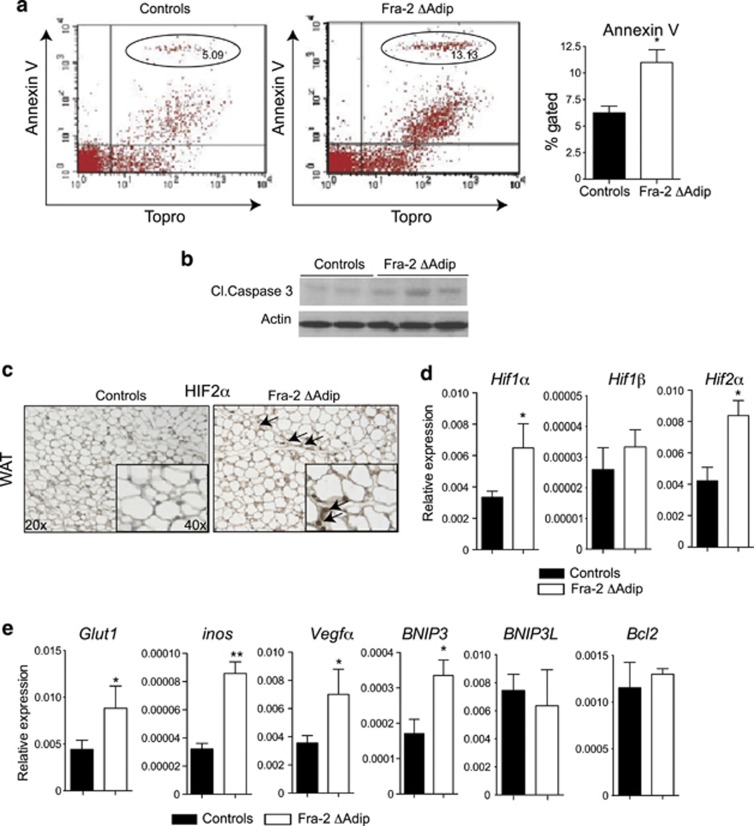
Figure 2.
Fra-2Δadip fat pads are hypoxic and show higher apoptosis rates. (a) Quantification of apoptotic cells by FACS in perigonadal fat from Fra-2Δadip and littermate control mice at 6 weeks after tamoxifen injection (n=4). (b) Western blot for cleaved caspase-3 in perigonadal fat from Fra-2Δadip and littermate control mice at 6 weeks after tamoxifen injection. (c) HIF2α staining in Fra-2Δadip and littermate controls fat pad at 6 weeks after tamoxifen injection. Magnification × 20, insert × 40. Black arrows indicate HIF-positive cells. (d) Real-time PCR analyses of HIF1α, HIF1β and HIF2α in Fra-2Δadip and littermate controls fat pad at 6 weeks after tamoxifen injection (n=4). (e) Real-time PCR analyses of HIFs target genes Glut1, Inos, Vegfα, BNIP3, BNIP3L and Bcl2 in Fra-2Δadip and littermate controls fat pad at 6 weeks after tamoxifen injection (n=4). Bars represent mean values±S.D. Statistical analyses: *P<0.05, **P<0.01

