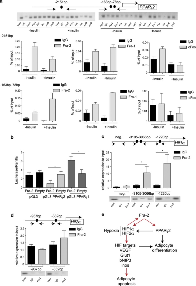
Figure 5.
PPARγ2 and HIF1α are transcriptional targets of Fra-2. (a) Chromatin immunoprecipitation (ChIP) for PPARγ2 promoter. Arrows indicate primers amplifying fragments for the TRE elements. Chromatin isolated from 3T3L1 cells with insulin treatment for 24 h was immunoprecipitated with IgG and AP-1 antibodies. Real-time PCR analyses and loading of the gel are shown (n=2). (b) Luciferase assay analyses of Fra-2 on PPARγ1 and PPARγ2 promoter; empty plasmids are shown for the control experiment. (c and d) ChIP for HIF1α (c) and HIF2α (d) promoters. Arrows indicate primers amplifying fragments for the TRE elements. Chromatin from 3T3L1 cells was immunoprecipitated with IgG and Fra-2 antibodies. Real-time PCR analyses and loading of the gel are shown (n=3). (e) Scheme of Fra-2 actions in adipocytes. Red arrows indicate findings from the manuscript and black arrows indicate knowledge from the literature. Bars represent mean values±S.D. Statistical analyses: *P<0.05

