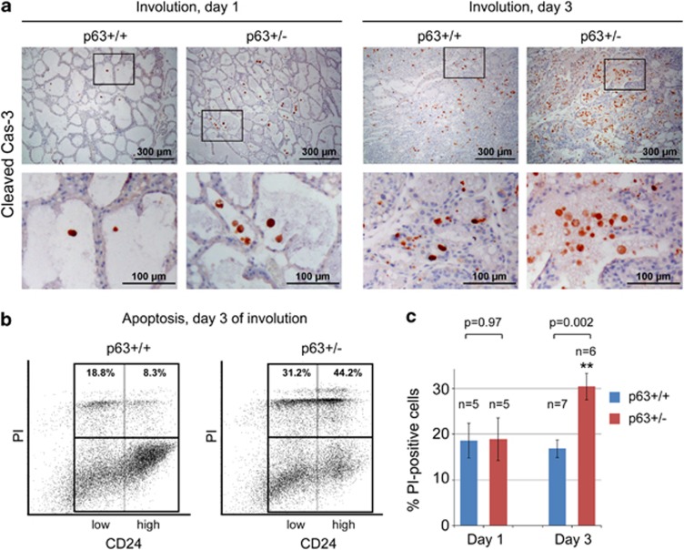
Figure 4.
Enhanced apoptosis in p63+/− mammary glands during early involution. (a) Immunostaining of cleaved caspase-3 in p63+/+ and p63+/− mammary glands at day 1 and day 3 of involution. Bottom images are enlargements of the boxed areas in the corresponding top images. One out of two independent experiments with similar results is shown. (b and c) FACS analysis of CD24positive cells stained for PI (propidium iodide) at day 1 and day 3 post involution. A representative analysis at day 3 (b) and quantification from four (day 1) and six (day 3) independent experiments (c) is shown. See also Supplementary Figure 3A for gating before the analysis shown here. Mean±S.E.M. N, number of animals analyzed, **P<0.01

