Figure 3.
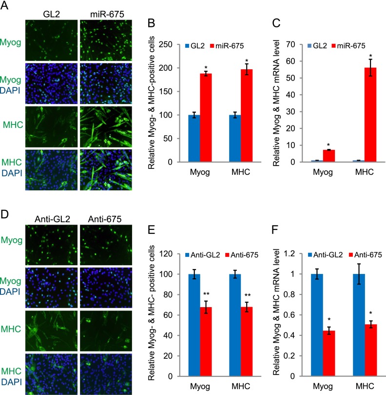
miR-675-3p and miR-675-5p promote muscle differentiation. (A) C2C12 myoblast cells in GM were transfected twice at 24-h intervals with GL2 control microRNA or miR-675-3p and miR-675-5p (miR-675). The cells were then transferred to DM and stained for myogenin at 32 h or MHC at 60 h. (Green) Myogenin or MHC; (blue) nuclei stained by DAPI. (B) Numbers of myogenin- and MHC-positive cells are presented relative to the GL2 control, which is set as 100%. Mean ± SEM of 10 random fields (see Supplemental Table 2 for details). (*) P < 0.001. (C) C2C12 myoblast cells were transfected as in A and kept in GM for an extra 24 h before harvesting to measure myogenin and MHC mRNA by qRT–PCR. Each value was normalized to GAPDH in the same sample and then again to the value in GL2-transfected cells. Mean ± SEM of three biological replicates. (*) P < 0.001. (D,E) 2′O-methyl antisense oligonucleotides against GL2 (anti-GL2) or miR-675-3p and miR-675-5p (anti-675) were transfected as in A, and the cells were stained for myogenin at 32 h or MHC at 60 h in DM. Data are presented as in B. (F) Measurement of myogenin and MHC mRNAs by qRT–PCR as in C. Mean ± SEM of three biological replicates are shown. (*) P < 0.001; (**) P < 0.005.

