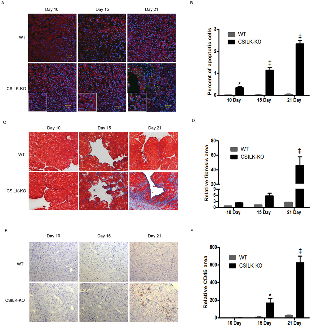
Figure 3. Increased apoptosis, fibrosis, and inflammation in CSILK-KO hearts.
Representative TUNEL-stained (A), Masson’s Trichrome-stained (C) or CD45-stained (E) cryosections of the heart from CSILK-KO mice and littermate controls at the indicated ages are shown, n=5 for each. Corresponding quantitative data are shown on the right panel, *p < 0.05, ‡ p < 0.001 vs. WT at each time point, n=5 (B, D, F).

