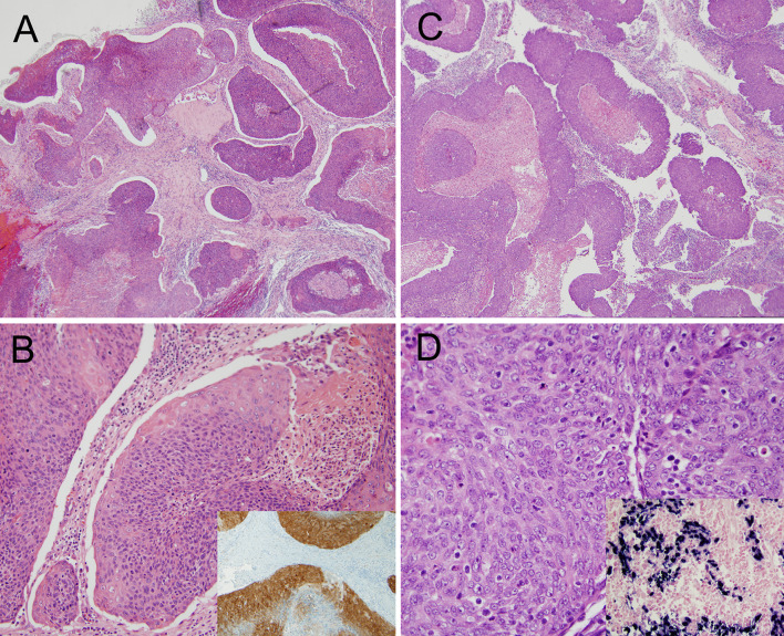
Fig. 5.
Nasopharyngeal carcinoma. a, b An HPV-related nasopharyngeal carcinoma showing ribbons of blue tumor with smooth edges and central necrosis. The inset shows positive p16 expression (a, ×4 magnification; b, ×20 magnification; b, inset ×10). c, d An EBV-related nasopharyngeal carcinoma also showing a ribbony appearance with smooth-edged nests and oval nuclei with vesicular chromatin and relatively inconspicuous nucleoli. The inset shows positivity for EBV early RNA by in situ hybridization. (c, ×4; d, ×40; d, inset ×20) (Images courtesy of Simion Chiosea M.D., University of Pittsburgh Medical Center)

