
Figure 2.
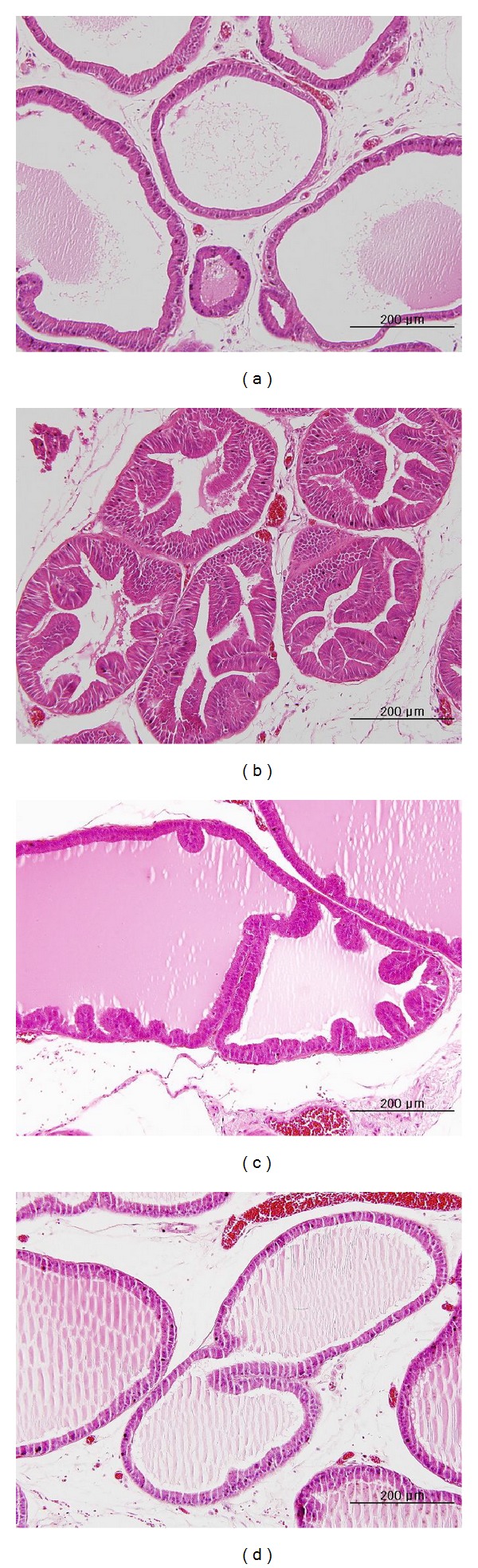
Histological analysis of prostate specimens taken from each experimental group. (a) Secretory luminal cells were lined with a single layer of low columnar epithelium and the acini were filled with pale eosinophilic materials (H & E stain, ×200). (b) The epithelial cells in glands were arranged as several uneven layers and the gland was excessively developed (H & E stain, ×200). (c) In comparison to (b), the proliferation of columnar epithelial cells in the BPH + SE1 group was restricted, and the development of the gland was limited (H & E stain, ×200). (d) It is difficult to find a difference between (d) and (a), except for the multiple layers of columnar epithelial cells in some parts of the gland in (d). H & E: hematoxylin and eosin; (a) control; (b) BPH; (c) BPH + SE1; (d) BPH + SE2.