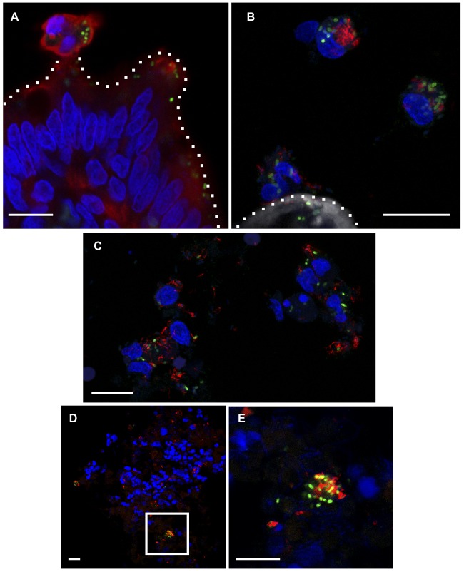
FIG 2 .
PinvF-positive, flagellated S. Typhimurium bacteria are associated with host cells proximal to villus tips and in the lumen. Ligated jejunal-ileal loops were infected with S. Typhimurium cells harboring PinvF-GFP[LVA] for 2 h. (A) Confocal image showing an extruded epithelial cell (cytokeratin 8 positive) containing numerous PinvF-GFP-positive bacteria. Green, anti-GFP; red, anti-cytokeratin 8; blue, DNA. (B) Confocal image showing several bovine cells adjacent to a villus tip containing a significant burden of flagellin-positive and PinvF-GFP positive Salmonella cells. Green, anti-GFP; red, anti-FliC; shades of gray, phalloidin for actin; blue, DNA. For A and B, dotted lines indicate the apical surface of the villus tip. (C) Confocal image of cells adjacent to villus tips revealing flagellin-positive and PinvF-GFP-positive bacteria intimately associated with bovine cells sloughed from the tissue. Green, anti-GFP; red, anti-FliC; blue, DNA. (D) Confocal image of luminal fluid content showing PinvF-GFP-positive, flagellated Salmonella bacteria found in clusters within a heterogeneous milieu of eukaryotic and prokaryotic cells. Green, anti-GFP; red, anti-FliC; blue, DNA. (E) Enlarged inset from panel D. Bars, 10 µm.

