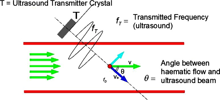
Fig. 2.
Schema justifying different Doppler signals occurring during THD as related to arterial blood flow. According to this physical law, the intensity of the Doppler signal is the inverse of the cosine of the angle between the ultrasound waves and blood flow. The more perpendicular the blood flow to the ultrasound waves (i.e., artery into the perirectal tissue or submucosa) the higher the Doppler signal; the more parallel the flow (i.e., artery perforating the rectal muscle) the lower the signal

