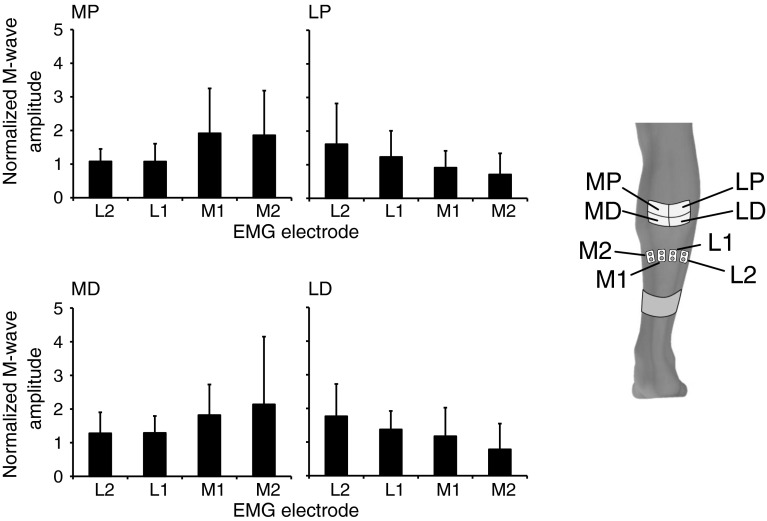
Fig. 7.
Grouped data of the normalized amplitude of the M-waves from each of the four muscle portions dependent on the location of the stimulation during SDSS (data normalized to SES). For SDSS electrodes, MP medial proximal; LP lateral proximal; MD medial distal; LD lateral distal. EMG electrodes placed over the m. soleus from medial to lateral portions as M2, M1, L1, and L2

