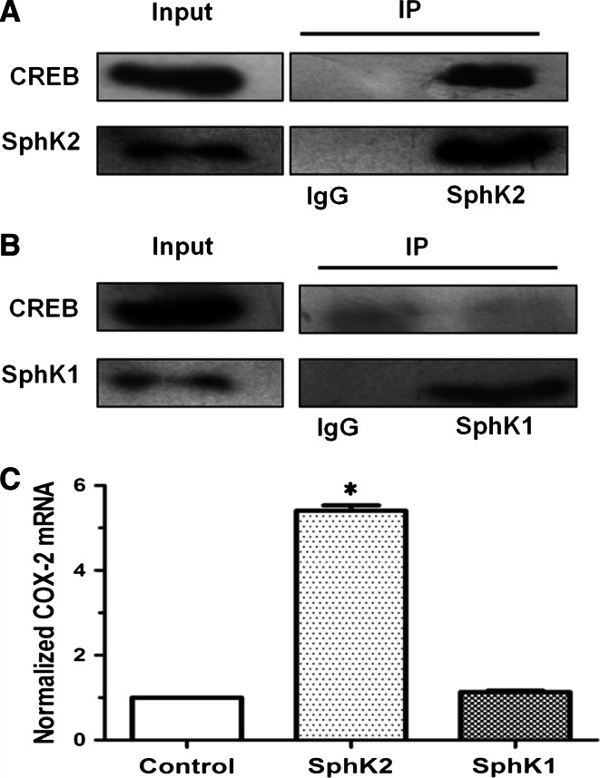
Fig. 8.
SphK-2 interacts with CREB. Cells were pulled down as described under experimental procedures. HUVECs were incubated with anti-SphK-2 or IgG antibodies coupled to agarose beads.Bound proteins were analyzed by Western blotting with anti-CREB and SphK-2 antibodies. 10 μg of total cell lysates from each sample were also analyzed by immunoblotting with anti-CREB and SphK-2 antibodies as inputs (a). HUVECs were incubated with anti-SphK-1 or IgG antibodies coupled to agarose beads. Bound proteins were analyzed by Western blotting with anti-CREB and SphK-1 antibodies. 10 μg of total cell lysates from each sample were also analyzed by immunoblotting with anti-P-CREB and SphK-1 antibodies as inputs (b). HUVEC cells incubated with anti-SphK1, or SphK2 antibodies coupled to agarose beads, then treated with vehicle or HDL (120 mg/ml) for 20 min and mRNA levels of COX-2 were determined by qPCR and normalized to GAPDH. c Results from at least three independent experiments conducted in triplicate are shown. *P < 0.05, versus cells treated with SphK-1 antibodies group

