Fig. 3.
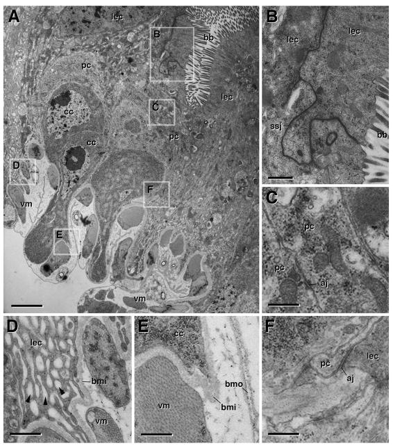
Ultrastructure of the metamorphosing midgut. White Prepupa. A: Cross section of wall of white pre-pupal midgut at low magnification. Boxed areas indicate regions shown at higher magnifications in panels B–F. B: Enterocytes of larval midgut (lec) with apical brush border (bb) and smooth septate junctions (ssj). C: peripheral cells (pc), stretching out to become transient pupal midgut, form leaflets interconnected by spot adherens junctions (aj). D: Basal part of larval enterocyte, containing numerous membrane invaginations (“labyrinth”; arrowheads). Inner layer of basement membrane (bmi) separates larval midgut cells from visceral muscle (vm). E: Basal process of central cell (cc) separated from visceral muscle by inner layer of basement membrane. Outer layer of basement membrane (bmo) covers visceral muscle. F: Spot adherens junctions (aj) link larval enterocytes and peripheral cells. aj adherens junction, bb brush border, cc central cells, bmi inner layer of basement membrane, bmo outer layer of basement membrane, lec larval enterocyte, pc peripheral cell, ssj smooth septate junction, vm visceral muscle. Bars: 2μm (A); 0.5μm (B); 0.4μm (C–F)

