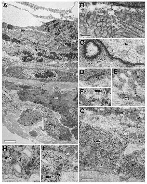
Fig. 4.
Ultrastructure of metamorphosing midgut, 6h pupa. A: Cross section showing degenerating larval gut (lmg), pupal midgut (pmg), adult midgut (amg), and visceral muscle (vm). Microvillar brush border (bb) is still visible in larval midgut. Mitochondria are fragmenting (arrowheads). B: Brush border of larval midgut at higher magnification. C, D: Septate junction (sj) between enterocytes of larval midgut (lec). E: Spot adherens junction (aj) between larval enterocyte and pupal midgut cell (pmg). F: Spot adherens junction between leaflets of pupal midgut. G–I: Cross section of basal part of larval midgut, pupal midgut, and adult midgut. Note that the labyrinth of basal membrane invaginations in larval enterocytes (see Fig. 3D) is all but gone; arrowhead in (G) points at one of the remnants of these invaginations. Adult midgut cells are connected basally by spot adherens junctions (aj; see higher magnification in H). Spot adherens junctions also connect adult to pupal midgut cells (I). Microvilli or septate junctions have not formed yet in adult midgut or pupal midgut. aj adherens junction, amg adult midgut, bb brush border, bmi inner layer of basement membrane, lec larval enterocyte, pmg pupal midgut, sj septate junction, vm visceral muscle. Bars: 2μm (A); 0.2μm (B–F; H, I); 1μm (G)

