Fig. 5.

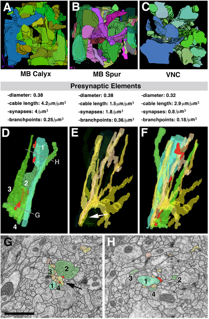
Structural network properties in Drosophila brain neuropile. Panels of the top row (A-C) show 3D digital models of presynaptic varicose/globular neurites from three different micro-volumes. A, B: micro-volumes from calyx (A; input region) and spur (B; output region) of mushroom body. In both micro-volumes, branches of terminal axonal neurites follow all directions. C: micro-volume from dorso-lateral neuropile of ventral nerve cord. All neurites are oriented predominantly along longitudinal axis. Shown below each panel are some core parameters of neurite profiles seen in micro-volumes. Note that average diameter of presynaptic elements (varicose and globular neurites) are very similar between the different regions. Density of presynaptic sites and branch points is significantly lower in VNC compared to mushroom body. D-H: Typical trajectories of presynaptic and postsynaptic terminal branches. D shows 3D digital models of four neighboring presynaptic neurites (1–4). Neurite 1 has a varicosity near top of panel (arrow); varicosities of the other neurites are more basally. E: bundle of dendritiform neurites extending in vicinity of terminal axons shown in D. F: terminal axons and dendrites shown together. G, H: EM sections close to top and bottom of VNC microvolume (levels of section shown in D). Profiles corresponding to the elements shown in models D-F are shaded in corresponding colors. As shown here, groups of dendritiform neurites (typically ranging between 6 and 10) form tight bundles in between adjacent preterminal axons (arrow in E, G). After forming synaptic contacts, dendritiform neurites typically splay apart (E) to then regroup with other dendrites in different configurations.
Bar: 1µm
