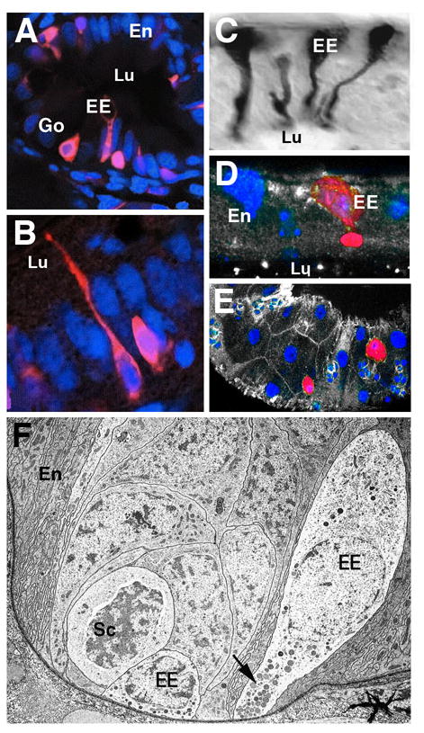
Fig.1.

Enteroendocrine cells in the vertebrate and Drosophila intestine. A, B: Five day zebrafish posterior intestine (from Crosnier et al., 2005). Enteroendocrine cells (EE) are labeled by monoclonal antibody 2F11 (red; nuclei of all cells labeled by TOPRO-3 in blue) exhibit an elongated, neuron-like shape, with a basal cell body and a slender apical process integrated into the enterocyte layer (En) and contacting the gut lumen (Lu). Exocrine goblet cells (Go) are also labeled. C: Endocrine cells in locust midgut, labeled by antibody against locust tachkinin-related peptide (from Winther and Nässel, 2001). Note characteristic shape and position of cells, resembling vertebrate enteroendocrine cells. D: Cross section of Drosophila adult midgut epithelium. Enteroendocrine cell labeled by anti-Tachykinin antibody (red). Cell nuclei labeled with Sytox (blue). As in locusts, endocrine cell body is located basally and possesses a club-shaped apical protrusion. E: Tangential section of Drosophila adult midgut epithelium, showing scattered distribution of tachykinin-positive endocrine cells (red). F: Electron micrograph of basal portion of midgut epithelium, showing enteroendocrine cells (EE) in close spatial association with proliferating stem cell “nests” (Sc; from Lehane, 1998). Note dense-core vesicles near basal membrane of endocrine cells (arrow).
