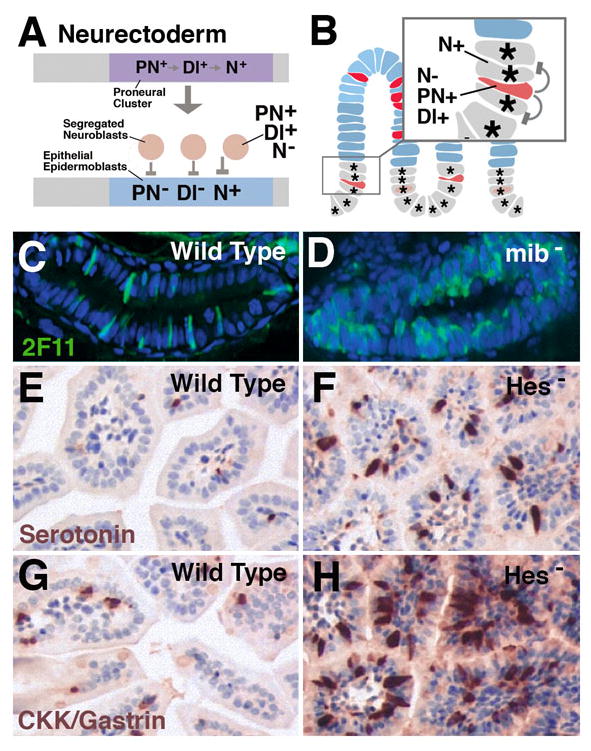
Fig.3.

Notch signaling controls the ratio of enteroendocrine cells and enterocytes in vertebrate intestinal epithelium. A: Canonical role of Notch signaling in proneural clusters of Drosophila neurectoderm. From an initially homogenous cell population forming the proneural cluster, two subpopulations are selected. Cells that upregulate Delta (Dl+) and proneural genes (PN+) and downregulate Notch activity (N-) become neuroblasts; the remainder upregulate Notch (N+) which suppresses proneural genes and Delta (PN-; Dl-, respectively. B: Intestinal progenitors can be compared to proneural cluster(s). Cells that upregulate Delta and proneural genes and downregulate Notch give rise to endocrine cells (postmitotic or progenitors); these cells activate Notch activity in the adjacent cells, which thereby are inhibited (‘lateral inhibition’) to adopt an endocrine fate, and become enterocytes and/or continue to proliferate. C: Wild type zebrafish larval gut in which secretory cells are labeled with antibody 2F11. D: In mindbomb (mib) mutant (elimination of Delta function), the majority of gut cells develops as secretory cells (endocrine and exocrine; from Crosnier et al., 2005). E, G: Wild type mouse intestinal epithelium in which endocrine cells expressing serotonin (E) or CKK/gastrin (G) are labeled. Both types of cells are increased in number in mutants where the Hes-1 gene (loss of Notch signaling activity) is impaired (F, H; from Jensen et al., 2000).
