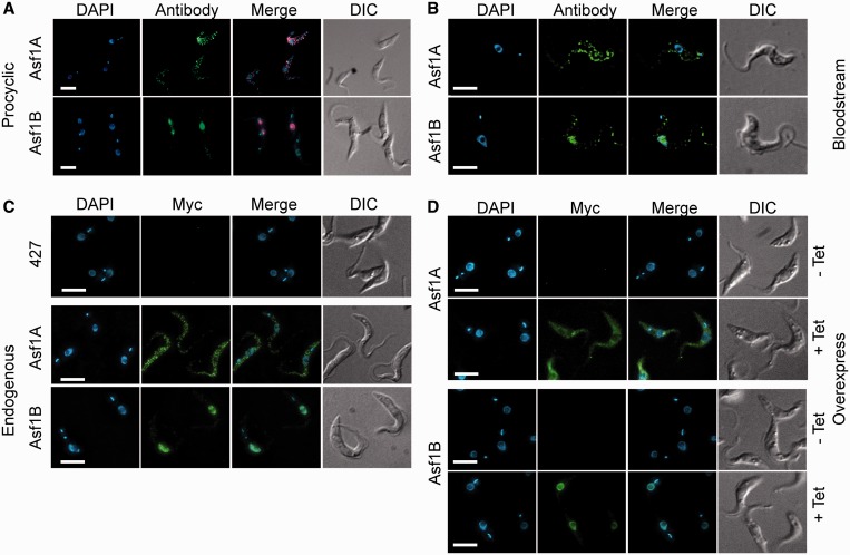
Figure 2.
T. brucei Asf1A is nuclear, whereas Asf1B is predominantly cytosolic. Panels (A) and (B) show IFA of procyclic and bloodstream T. brucei, respectively, using specific anti-Asf1A and anti-Asf1B antibodies (antibody), DAPI to stain DNA, merged and DIC images. Panel (C) shows IFA using anti-Myc monoclonal antibody, DAPI staining, merged images and DIC of wild-type procyclics (427) or procyclics expressing the endogenously 12×Myc-tagged versions of Asf1 or Asf1B. Panel (D) shows representative images of procyclics containing the plasmids pRP-Asf1A and pRP-Asf1B, which enable induced overexpression of proteins tagged with a 6×Myc peptide at the N-terminus. The analysis was performed in cells after 48 h without (−Tet) or with tetracycline-induced (+Tet) protein overexpression. Bars = 5 μm.

