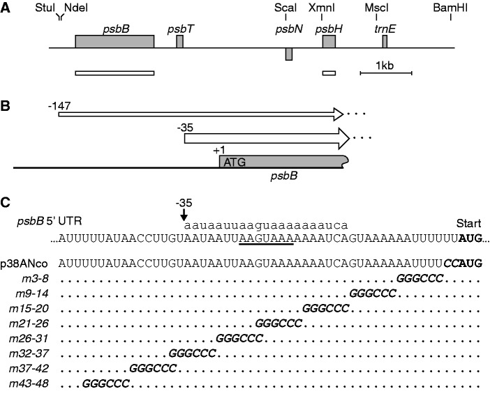
Figure 2.
Mutational analysis of the psbB 5' UTR. (A) Map of the psbB/psbT/psbH locus. The gray bars represent the open reading frames of psbB, psbT and psbH, and below the line of the putative psbN, which would be transcribed from the opposite strand. Restriction sites relevant to this work are indicated at the top, and the probes used for RNA blot hybridization are shown below as white bars. (B) The 5' UTRs of the psbB transcripts. The major form of the psbB 5' UTR begins at −35 relative to the start codon. A minor form, which is two orders of magnitude less abundant, begins at −147 (19). (C) Linker-scan mutagenesis of the psbB 5' UTR. The sequence of the WT is shown at the top with the translation start codon of psbB highlighted in bold and the S-box underlined. The corresponding sRNA (psbB 5') is shown in lowercase above the sequence. The black arrow shows the positions of the major 5'-end at −35. The sequences of the chloroplast transformation vector p38ANco and of the eight linker-scan mutants are aligned below, with the ApaI sites in italics and with dots representing unchanged bases.

