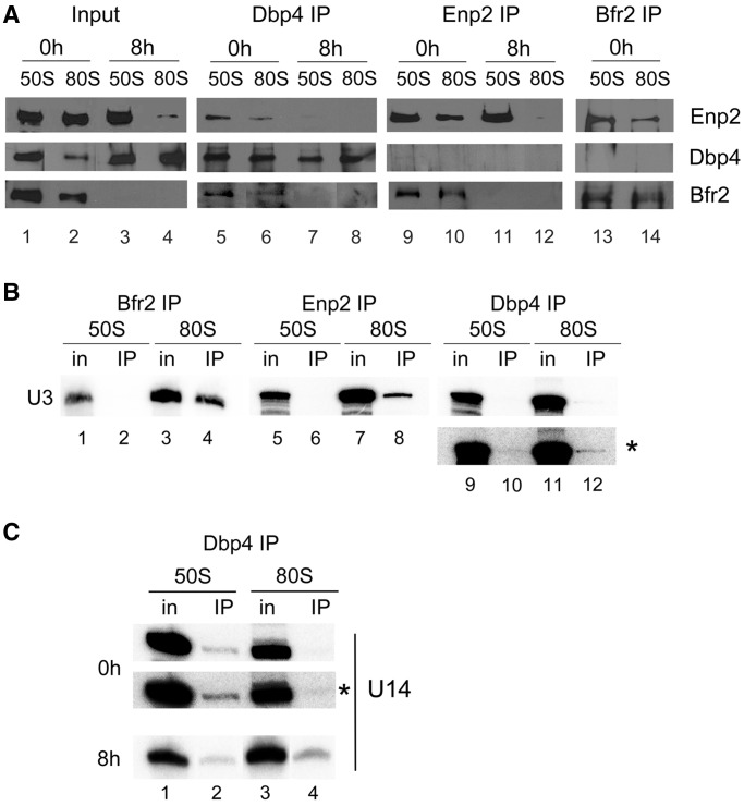
Figure 8.
Association of Bfr2, Dbp4 and Enp2 in complexes of ‘50S’ and ‘80S’ isolated from sucrose gradients. (A) Cellular extracts obtained from undepleted and Bfr2-depleted cells were fractionated on sucrose gradients as in Figure 7A, and two series of inputs (In) were prepared for IPs: pooled fractions 3–5 correspond to the ‘50S’ complex and fractions 7–8 are the ‘80S’ complex. IPs were done with anti-Dbp4 (lanes 5–8), anti-myc (lanes 9–12), and anti-HA antibodies (lanes 13 and 14). Western blot analyses were carried out using the same antibodies to detect the presence of Enp2 (myc), Bfr2 (HA) and Dbp4. Input lanes correspond to 12% of pooled fractions. (B) Gradient fractions were prepared from undepleted cells and IPs were done as in Figure 8A, except that RNAs were extracted and subjected to northern hybridization with a radiolabeled oligonucleotide complementary to the U3 snoRNA. Inputs (In) correspond to 10%. The asterisk indicates the overexposed blot of Dbp4 IP. (C) IPs were done with anti-Dbp4 antibodies as in Figure 8B, except that northern hybridization was carried out with a radiolabeled oligonucleotide complementary to the U14 snoRNA.

