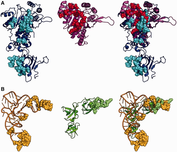
Figure 1.
Contact surface in EF-P/PoxA and tRNAAsp/AspRS complexes. The image shows a comparison of the polar contacts between EF-P and PoxA with polar contacts between tRNAAsp and AspRS. Image corresponds to a superposition of AspRS and PoxA from pdb files 1asy (chains A and R) and 3a5z (chains C and D). AspRS is in blue, tRNAAsp in orange, PoxA in magenta and EFP in green. Amino acids and nucleotides making polar contacts are marked in balls with the following colors: AspRS in light blue, tRNAAsp in light orange, EFP in light green and PoxA in red. (A) Enzymes. (B) Substrates.

