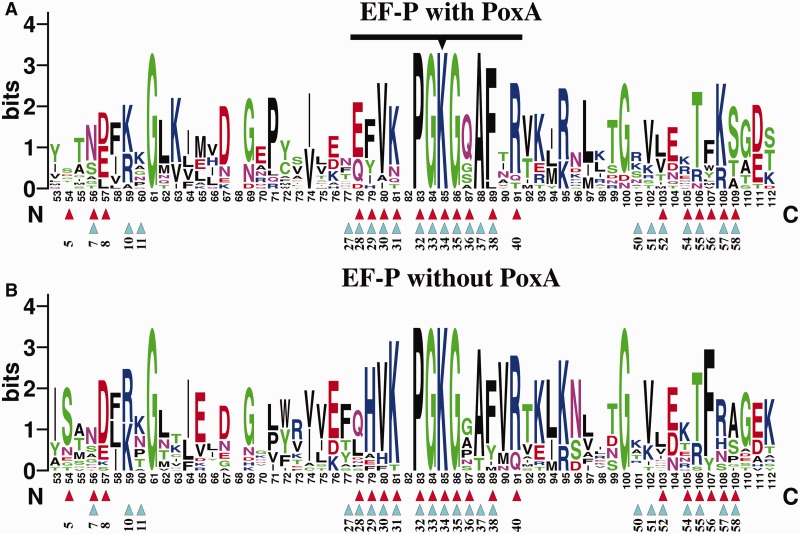
Figure 4.
Conservation of EF-P residues involved in PoxA contacts. WebLogo representation of a fragment of the alignment of diverse EF-Ps that contain a lysine in the equivalent to the modification position. The top WebLogo (A) corresponds to an alignment of 14 EF-P sequences from organisms that have a poxA gene encoded in their genome. The bottom WebLogo (B) corresponds to an alignment of 17 EF-P sequences from organisms that do not have a poxA gene. The acceptor loop is marked with a black line, with the modification position highlighted with a triangle. Contacts to PoxA (based on pdb 3a5z) are marked with red triangles and contacts to the ribosome (pdb 3huw and 3hux) are indicated with cyan triangles. Numbering of the WebLogo positions corresponds to the full alignment, and corresponding positions on EF-P from E. coli are indicated below the triangles (for full alignments see Supplementary Figure S4).

