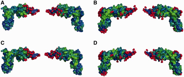
Figure 5.
EF-P contacts with PoxA and the ribosome. Figure shows EF-P structures from pdb 3a5a-D (A and C) or 3huw-V (B and D). Atoms from PoxA (C and E) or the ribosome and tRNA (D and F) that contact EF-P are highlighted in red. Variable positions of the alignments (Figure 4 and Supplementary Figure S4) are highlighted on the EF-P surface for organisms with (C and D) or without (E and F) poxA. Variable positions are in green and non-variable are in blue.

