
Figure 1.
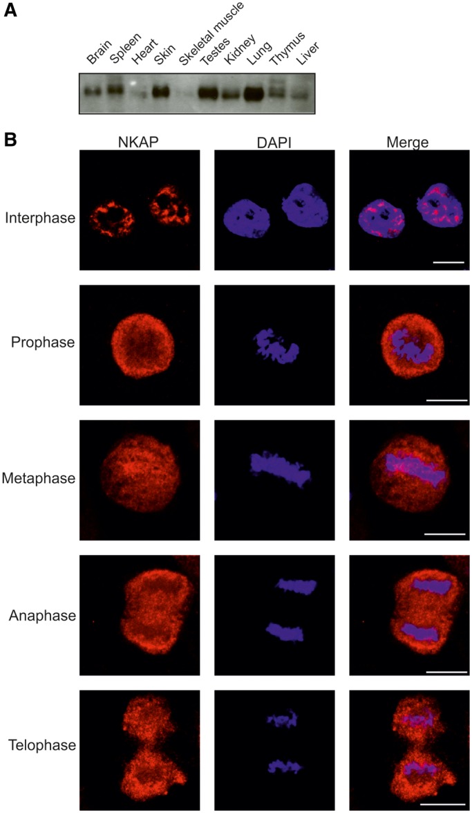
Expression and localization of NKAP. (A) Presence of NKAP in mouse tissues. Homogenates of adult mouse tissues as indicated were separated by SDS-PAGE (12% acrylamide) and the corresponding blot probed with NKAP-specific mAb K85-80-5. (B) Localization of NKAP during mitosis. HeLa cells were synchronized using nocodazole to block progression of the cell cycle and then released and fixed using 4% paraformaldehyde (PFA). Cells were stained with mAb K85-80-5, and nuclei (blue) were stained with 4′,6-diamidino-2-phenylindole (DAPI). The phases of the cell cycle are indicated. Bar, 10 µm.