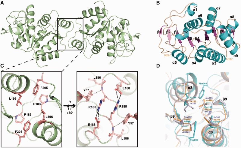
Figure 2.
Structures of apo-HpDprA(5-225). (A) The dimer of HpDprA(5-225) colored in pale green. (B) The secondary structure elements of the monomer are labeled in different colors. (C) The dimerization interface of HpDprA(5-225). Residues involved in dimerization are shown in stick models. (D) The superimpositions of the hydrophobic cores. The hydrophobic residues are shown as stick models with those of RpDprA in orange, of SpDprA in white and of HpDprA in cyan. These residues are also marked as blue stars in Figure 1C.

