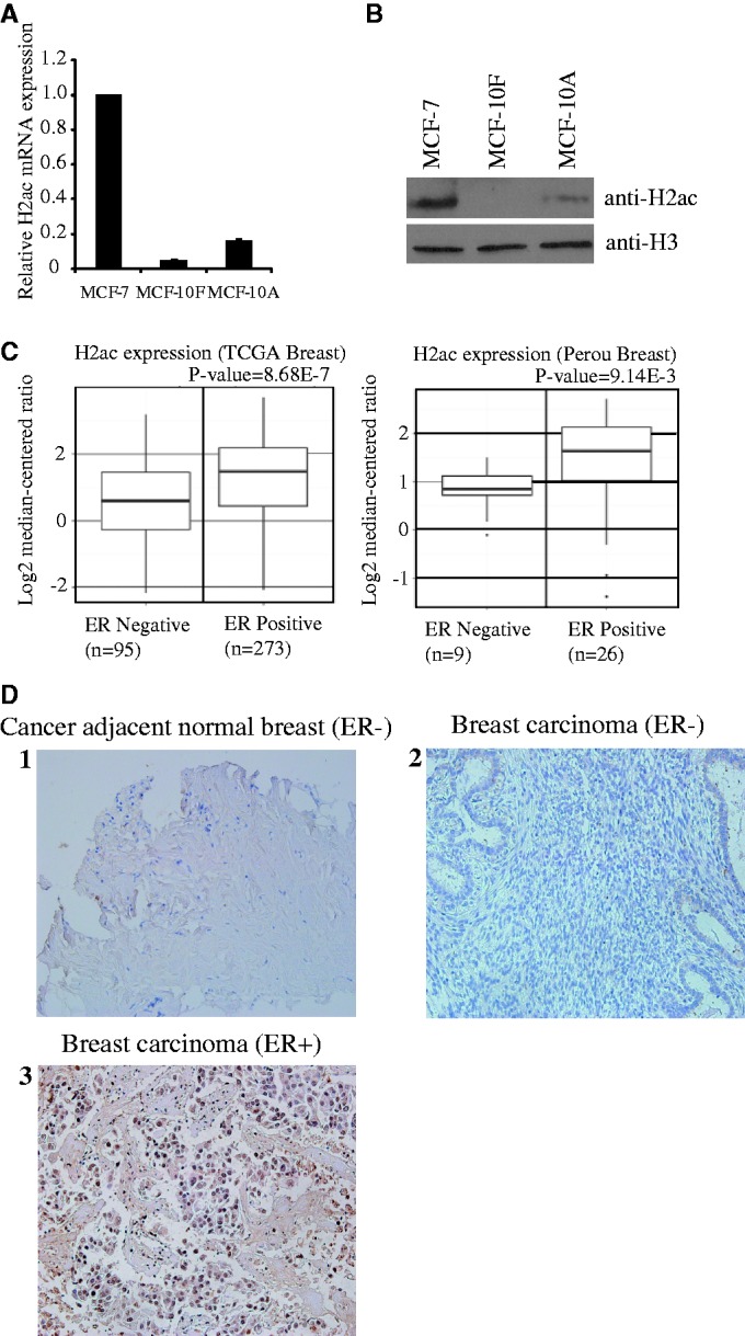
Figure 1.

H2ac is overexpressed in MCF-7 cells and is associated with estrogen-receptor positive breast carcinoma. (A) mRNA expression levels of H2ac were determined by quantitative RT-PCR and normalized against 18s rRNA. (B) Western blotting of histone extracts prepared from MCF-7, MCF-10F and MCF-10A cells using the antibodies shown in the right panel. (C) Increased expression of H2ac mRNA in ER+ breast carcinomas from two independent data sets for human breast cancers. Data and statistics were obtained from the Oncomine database (14,15). (D) IHC analysis show H2ac protein expression in carcinoma samples of ER+ breast cancer patients. IHC was used to examine H2ac expression in TMAs and compare it with that of human ER− adjacent normal breast tissues, human malignant ER− breast cancer tissues and human malignant ER+ breast cancer tissues. Counterstaining used hematoxylin. Representatives of normal adjacent breast tissue (1), ER− malignant breast tissue (2) and ER+ malignant breast tissue (3) are shown.
