
Figure 1.
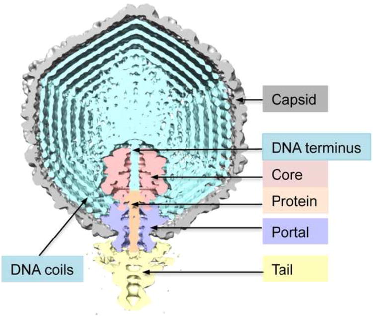
Segmented cutaway model of the T7 virion, taken from a cryo-EM reconstruction in which the only symmetry imposed and exploited was C5 symmetry around the portal axis. The various components are color-coded as shown. The region along the portal axis extending from the start of the portal is occupied by positive density. Based on bubblegram imaging (Results and Discussion), we identify the portal-proximal part (light brown) as protein and the portal-distal part as the “left” end of the genome, viz. “DNA terminus”. The virion has 6 tail-fibers distributed around the proximal portion of the tail but they are not seen in this reconstruction.