
Figure 2.
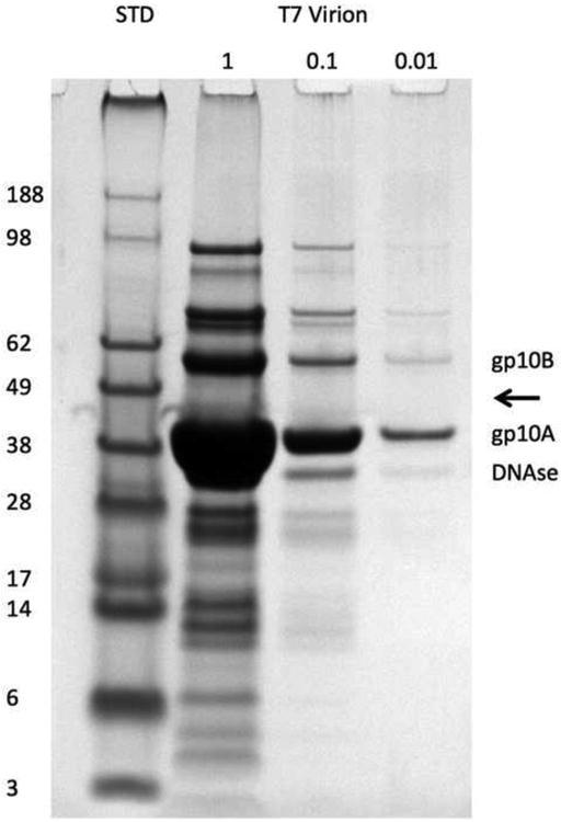
Proteins from purified T7 virions separated by reducing SDS-PAGE and stained with Coomassie Blue. The first lane (STD, standards) shows reference proteins with their molecular masses in kDa indicated on the left. The next three lanes show loadings of T7 proteins at serial 10-fold dilutions. The major and minor capsid proteins, gp10A and gp10B, are labeled, as is DNAse 1 which was added to digest viral DNA. The expected location of the scaffolding protein gp9 (Roeder and Sadowski, 1977) is indicated by an arrow.