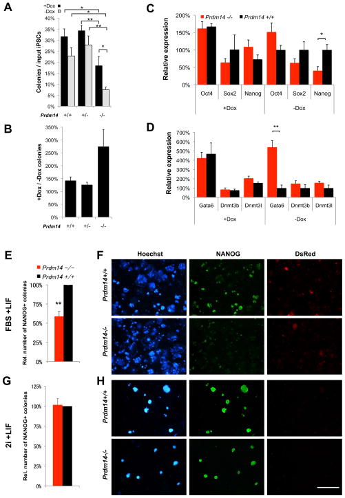
Figure 5. The self-renewal defect of Prdm14−/− iPSCs is partially rescued by maintained exogenous pluripotency factor expression or by derivation and culture in 2i+LIF.
(A, B) Replating efficiency of iPSCs depending on exogenous pluripotency factor expression. Primary iPSCs were derived in 11 days with Dox before reseeding and culture for 14 days with (+Dox) or without (−Dox) induction of the lentiviral reprogramming cassette. Replating efficiency was measured as AP-positive colonies per seeded input iPSCs (A). * P<0.05, ** P<0.01 2-way ANOVA with Bonferroni post-tests to compare individual means. In (B), the relative colony number increase of continuous Dox-treatment over Dox-withdrawal is shown. Error bars = SEM.
(C, D) Quanititative (q)PCR measurements of gene expression (C) in Prdm14+/+ (black) and Prdm14−/− (red) iPSCs cultured with (+Dox) or without (−Dox) exogenous OKSM factors. (C) Endogenous pluripotency gene expression. * P=0.034 two-tailed Student’s t-test. (D) Gata6 (endodermal differentiation marker) and de novo DNA-methyltransferase expression. ** P=0.00097. Error bars = SEM.
(E–H) Comparison of derivation efficiency of Prdm14+/+ vs. Prdm14−/− iPSCs under FBS+LIF (E, F) or 2i+LIF conditions (G, H) using retroviral vectors. (E, G) Relative number of NANOG+ colonies from reprogrammed Prdm14−/− cells (red) compared to Prdm14+/+ (black, set to 100%). ** P=0.008 two-tailed Student’s t-test. Error bars = SEM. (F, H) NANOG-activation and silencing of retroviral vectors (DsRed-negative) in iPSC-colonies as indicators of successful reprogramming (Hoechst = DNA). Scale bar = 4 mm.

